 ムービー
ムービー コントローラー


+ データを開く

- 基本情報
基本情報
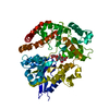
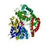
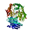
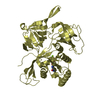
| 登録情報 | データベース: PDB / ID: 4zlt | |||||||||||||||
|---|---|---|---|---|---|---|---|---|---|---|---|---|---|---|---|---|
| タイトル | Crystal structure of viral chemokine binding protein R17 in complex with CCL3 | |||||||||||||||
 要素 要素 |
| |||||||||||||||
 キーワード キーワード | Chemokine binding protein/Chemokine / RHVP chemokine binding protein in complex with chemokine CCL3 / Chemokine binding protein-Chemokine complex | |||||||||||||||
| 機能・相同性 |  機能・相同性情報 機能・相同性情報Chemokine receptors bind chemokines / lymphocyte chemotaxis / granulocyte chemotaxis / positive regulation of natural killer cell chemotaxis / astrocyte cell migration / eosinophil degranulation / regulation of sensory perception of pain / signaling / cell activation / T cell chemotaxis ...Chemokine receptors bind chemokines / lymphocyte chemotaxis / granulocyte chemotaxis / positive regulation of natural killer cell chemotaxis / astrocyte cell migration / eosinophil degranulation / regulation of sensory perception of pain / signaling / cell activation / T cell chemotaxis / eosinophil chemotaxis / positive regulation of calcium ion transport / response to cholesterol / positive regulation of osteoclast differentiation / chemokine activity / release of sequestered calcium ion into cytosol by sarcoplasmic reticulum / phospholipase activator activity / chemoattractant activity / negative regulation of osteoclast differentiation / exocytosis / macrophage chemotaxis / monocyte chemotaxis / host-mediated suppression of viral transcription / neutrophil chemotaxis / cytoskeleton organization / positive regulation of calcium-mediated signaling / positive regulation of interleukin-1 beta production / calcium-mediated signaling / response to toxic substance / chemotaxis / intracellular calcium ion homeostasis / positive regulation of tumor necrosis factor production / positive regulation of inflammatory response / kinase activity / osteoblast differentiation / calcium ion transport / cell-cell signaling / regulation of cell shape / positive regulation of neuron apoptotic process / positive regulation of cytosolic calcium ion concentration / protein phosphorylation / positive regulation of ERK1 and ERK2 cascade / protein kinase activity / positive regulation of cell migration / response to xenobiotic stimulus / inflammatory response / negative regulation of gene expression / positive regulation of gene expression / positive regulation of transcription by RNA polymerase II / : / cytoplasm / cytosol 類似検索 - 分子機能 | |||||||||||||||
| 生物種 |  Cricetid herpesvirus 2 (ヘルペスウイルス) Cricetid herpesvirus 2 (ヘルペスウイルス) | |||||||||||||||
| 手法 |  X線回折 / シンクロトロン / 分子置換 / 解像度: 3 Å X線回折 / シンクロトロン / 分子置換 / 解像度: 3 Å | |||||||||||||||
 データ登録者 データ登録者 | Lubman, O.Y. / Fremont, D.H. | |||||||||||||||
| 資金援助 |  米国, 4件 米国, 4件
| |||||||||||||||
 引用 引用 | ジャーナル: Structure / 年: 2016 タイトル: Parallel Evolution of Chemokine Binding by Structurally Related Herpesvirus Decoy Receptors. 著者: Lubman, O.Y. / Fremont, D.H. | |||||||||||||||
| 履歴 |
|
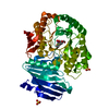
- 構造の表示
構造の表示
| 構造ビューア | 分子: MolmilJmol/JSmol |
|---|
- ダウンロードとリンク
-ダウンロード
| PDBx/mmCIF形式 | 4zlt.cif.gz | 187.1 KB | 表示 | PDBx/mmCIF形式 |
|---|---|---|---|---|
| PDB形式 | pdb4zlt.ent.gz | 148.9 KB | 表示 | PDB形式 |
| PDBx/mmJSON形式 | 4zlt.json.gz | ツリー表示 | PDBx/mmJSON形式 | |
| その他 |  その他のダウンロード その他のダウンロード |
-検証レポート
| アーカイブディレクトリ | https://data.pdbj.org/pub/pdb/validation_reports/zl/4zltftp://data.pdbj.org/pub/pdb/validation_reports/zl/4zlt | HTTPS FTP |
|---|
-関連構造データ


-リンク
 PDBj
PDBj
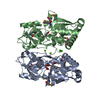
- 集合体
集合体
| 登録構造単位 | 
| ||||||||
|---|---|---|---|---|---|---|---|---|---|
| 1 | 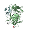
| ||||||||
| 2 | 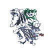
| ||||||||
| 単位格子 |
| ||||||||
| 詳細 | dimer according to multi-angle static light scattering |
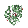
-要素
| #1: タンパク質 | 分子量: 47388.625 Da / 分子数: 2 / 変異: K333D, R335E, R336E, K337D / 由来タイプ: 組換発現 由来: (組換発現) Cricetid herpesvirus 2 (ヘルペスウイルス)遺伝子: RHVP-L.R17, RHVP.R17 / Cell (発現宿主): endothelial / 細胞株 (発現宿主): 293F / 発現宿主: Mammalian expression vector pBGSA (その他) / 参照: UniProt: E9M5R0 #2: タンパク質 | 分子量: 7982.065 Da / 分子数: 2 / 変異: D27A / 由来タイプ: 組換発現 / 由来: (組換発現)  #3: 糖 | ChemComp-NAG /   タイプ: D-saccharide, beta linking / 分子量: 221.208 Da / 分子数: 4 / 由来タイプ: 組換発現 / 式: C8H15NO6 タイプ: D-saccharide, beta linking / 分子量: 221.208 Da / 分子数: 4 / 由来タイプ: 組換発現 / 式: C8H15NO6Has protein modification | Y | |
|---|
-実験情報
-実験
| 実験 | 手法: X線回折 |
|---|
- 試料調製
試料調製
| 結晶 | マシュー密度: 2.57 Å3/Da / 溶媒含有率: 52.1 % |
|---|---|
| 結晶化 | 温度: 297 K / 手法: 蒸気拡散法, ハンギングドロップ法 / 詳細: 15-20% PEG 3350 0.2-0.4M MgFormate |
-データ収集
| 回折 | 平均測定温度: 100 K | |||||||||
|---|---|---|---|---|---|---|---|---|---|---|
| 放射光源 | 由来: シンクロトロン / サイト: ALS / ビームライン: 4.2.2 / 波長: 1 Å | |||||||||
| 検出器 | タイプ: NOIR-1 / 検出器: CCD / 日付: 2013年6月13日 | |||||||||
| 放射 | プロトコル: SINGLE WAVELENGTH / 単色(M)・ラウエ(L): M / 散乱光タイプ: x-ray | |||||||||
| 放射波長 | 波長: 1 Å / 相対比: 1 | |||||||||
| 反射 | 解像度: 2.761→50 Å / Num. obs: 26825 / % possible obs: 100 % / 冗長度: 4.7 % / Rsym value: 0.11 / Net I/σ(I): 7.4 | |||||||||
| 反射 シェル |
|
- 解析
解析
| ソフトウェア |
| |||||||||||||||||||||||||||||||||||||||||||||||||||||||||||||||||||||||||||||||||||||||||||
|---|---|---|---|---|---|---|---|---|---|---|---|---|---|---|---|---|---|---|---|---|---|---|---|---|---|---|---|---|---|---|---|---|---|---|---|---|---|---|---|---|---|---|---|---|---|---|---|---|---|---|---|---|---|---|---|---|---|---|---|---|---|---|---|---|---|---|---|---|---|---|---|---|---|---|---|---|---|---|---|---|---|---|---|---|---|---|---|---|---|---|---|---|
| 精密化 | 構造決定の手法: 分子置換 開始モデル: 4ZKQ and 2X6G 解像度: 3→49.247 Å / SU ML: 0.4 / 交差検証法: FREE R-VALUE / σ(F): 0 / 位相誤差: 27.94 / 立体化学のターゲット値: ML
| |||||||||||||||||||||||||||||||||||||||||||||||||||||||||||||||||||||||||||||||||||||||||||
| 溶媒の処理 | 減衰半径: 0.9 Å / VDWプローブ半径: 1.11 Å / 溶媒モデル: FLAT BULK SOLVENT MODEL | |||||||||||||||||||||||||||||||||||||||||||||||||||||||||||||||||||||||||||||||||||||||||||
| 精密化ステップ | サイクル: LAST / 解像度: 3→49.247 Å
| |||||||||||||||||||||||||||||||||||||||||||||||||||||||||||||||||||||||||||||||||||||||||||
| 拘束条件 |
| |||||||||||||||||||||||||||||||||||||||||||||||||||||||||||||||||||||||||||||||||||||||||||
| LS精密化 シェル |
|
