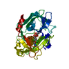
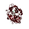
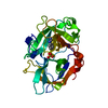
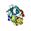
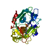
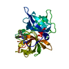
[English] 日本語
Yorodumi
- PDB-4zks: The crystal structure of upain-1-W3A in complex with inactive uPA... -

+
Open data


ID or keywords:

Loading...

-
Basic information

Entry
Database: PDB / ID: 4zks
TitleThe crystal structure of upain-1-W3A in complex with inactive uPA (uPA-S195A) at pH7.4
Components
  • Urokinase-type plasminogen activator
  • upain-1-W3A
KeywordsHYDROLASE/HYDROLASE INHIBITOR / Peptides inhibitor / uPA / serine protease / HYDROLASE-HYDROLASE INHIBITOR complex
Function / homology
Function and homology information


u-plasminogen activator / regulation of smooth muscle cell-matrix adhesion / urokinase plasminogen activator signaling pathway / regulation of plasminogen activation / regulation of fibrinolysis / regulation of wound healing / protein complex involved in cell-matrix adhesion / negative regulation of plasminogen activation / regulation of smooth muscle cell migration / regulation of signaling receptor activity ...u-plasminogen activator / regulation of smooth muscle cell-matrix adhesion / urokinase plasminogen activator signaling pathway / regulation of plasminogen activation / regulation of fibrinolysis / regulation of wound healing / protein complex involved in cell-matrix adhesion / negative regulation of plasminogen activation / regulation of smooth muscle cell migration / regulation of signaling receptor activity / serine-type endopeptidase complex / Dissolution of Fibrin Clot / smooth muscle cell migration / plasminogen activation / regulation of cell adhesion mediated by integrin / tertiary granule membrane / negative regulation of fibrinolysis / regulation of cell adhesion / specific granule membrane / serine protease inhibitor complex / fibrinolysis / chemotaxis / blood coagulation / regulation of cell population proliferation / response to hypoxia / positive regulation of cell migration / external side of plasma membrane / serine-type endopeptidase activity / focal adhesion / Neutrophil degranulation / cell surface / signal transduction / proteolysis / extracellular space / extracellular exosome / extracellular region / plasma membrane
Similarity search - Function
Kringle domain / Kringle / Kringle, conserved site / Kringle superfamily / Kringle domain signature. / Kringle domain profile. / Kringle domain / Kringle-like fold / EGF-like domain profile. / EGF-like domain signature 1. ...Kringle domain / Kringle / Kringle, conserved site / Kringle superfamily / Kringle domain signature. / Kringle domain profile. / Kringle domain / Kringle-like fold / EGF-like domain profile. / EGF-like domain signature 1. / EGF-like domain / Serine proteases, trypsin family, histidine active site / Serine proteases, trypsin family, serine active site / Peptidase S1A, chymotrypsin family / Serine proteases, trypsin family, histidine active site. / Serine proteases, trypsin domain profile. / Serine proteases, trypsin family, serine active site. / Trypsin-like serine protease / Serine proteases, trypsin domain / Trypsin / Trypsin-like serine proteases / Thrombin, subunit H / Peptidase S1, PA clan, chymotrypsin-like fold / Peptidase S1, PA clan / Beta Barrel / Mainly Beta
Similarity search - Domain/homology
Urokinase-type plasminogen activator
Similarity search - Component
Biological speciesHomo sapiens (human)
synthetic construct (others)
MethodX-RAY DIFFRACTION / SYNCHROTRON / Resolution: 1.85 Å
AuthorsJiang, L. / Andreasen, P.A. / Huang, M.
CitationJournal: To Be Published
Title: The crystal structure of mupain-1-IG in complex with murinised human uPA at pH7.4
Authors: Jiang, L. / Andreasen, P.A. / Huang, M.
History
DepositionApr 30, 2015Deposition site: RCSB / Processing site: PDBJ
Revision 1.0May 18, 2016Provider: repository / Type: Initial release

-
Structure visualization

Structure viewerMolecule:
MolmilJmol/JSmol

Downloads & links

-
Assembly

Deposited unit
U: Urokinase-type plasminogen activator
P: upain-1-W3A


Theoretical massNumber of molelcules
Total (without water)29,0622
Polymers29,0622
Non-polymers00
Water1,60389
1


  • Idetical with deposited unit
  • defined by author&software
TypeNameSymmetry operationNumber
identity operation1_555x,y,z1
Buried area1270 Å2
ΔGint-3 kcal/mol
Surface area11420 Å2
MethodPISA
Unit cell
Length a, b, c (Å)120.751, 120.751, 42.961
Angle α, β, γ (deg.)90.00, 90.00, 120.00
Int Tables number146
Space group name H-MH3

-
Components

#1: Protein Urokinase-type plasminogen activator / uPA


Mass: 27828.715 Da / Num. of mol.: 1 / Fragment: UNP residues 179-425 / Mutation: C122A, N145Q, S195A
Source method: isolated from a genetically manipulated source
Source: (gene. exp.) Homo sapiens (human) / Gene: PLAU / Production host: Komagataella pastoris (fungus) / Strain (production host): X33 / References: UniProt: P00749, u-plasminogen activator
#2: Protein/peptide upain-1-W3A


Mass: 1233.379 Da / Num. of mol.: 1 / Source method: obtained synthetically / Source: (synth.) synthetic construct (others)
#3: Water ChemComp-HOH / water


Mass: 18.015 Da / Num. of mol.: 89 / Source method: isolated from a natural source / Formula: H2O

-
Experimental details

-
Experiment

ExperimentMethod: X-RAY DIFFRACTION / Number of used crystals: 1

-
Sample preparation

CrystalDensity Matthews: 2.07 Å3/Da / Density % sol: 40.7 %
Crystal growTemperature: 298 K / Method: vapor diffusion, sitting drop / pH: 4.6
Details: 50 mM sodium citrate pH 4.6, 2.0 M ammonium sulfate supplemented with 5% PEG400

-
Data collection

DiffractionMean temperature: 100 K
Diffraction sourceSource: SYNCHROTRON / Site: SSRF / Beamline: BL17U / Wavelength: 0.97 Å
DetectorType: ADSC QUANTUM 315r / Detector: CCD / Date: Mar 31, 2014
RadiationProtocol: SINGLE WAVELENGTH / Monochromatic (M) / Laue (L): M / Scattering type: x-ray
Radiation wavelengthWavelength: 0.97 Å / Relative weight: 1
ReflectionResolution: 1.85→60 Å / Num. obs: 19848 / % possible obs: 99.8 % / Redundancy: 3.8 % / Net I/σ(I): 22.3

-
Processing

Software
NameVersionClassification
REFMAC5.7.0029refinement
HKL-2000data scaling
Cootmodel building
RefinementResolution: 1.85→33.22 Å / Cor.coef. Fo:Fc: 0.954 / Cor.coef. Fo:Fc free: 0.926 / SU B: 3.572 / SU ML: 0.109 / Cross valid method: THROUGHOUT / ESU R: 0.167 / ESU R Free: 0.155 / Stereochemistry target values: MAXIMUM LIKELIHOOD / Details: HYDROGENS HAVE BEEN ADDED IN THE RIDING POSITIONS
RfactorNum. reflection% reflectionSelection details
Rfree0.24457 1017 5.1 %RANDOM
Rwork0.19599 ---
obs0.19846 18829 99.61 %-
Solvent computationIon probe radii: 0.8 Å / Shrinkage radii: 0.8 Å / VDW probe radii: 1.2 Å / Solvent model: MASK
Displacement parametersBiso mean: 31.097 Å2
Baniso -1Baniso -2Baniso -3
1--0.41 Å2-0.41 Å20 Å2
2---0.41 Å2-0 Å2
3---1.35 Å2
Refinement stepCycle: 1 / Resolution: 1.85→33.22 Å
ProteinNucleic acidLigandSolventTotal
Num. atoms2025 0 0 89 2114
Refine LS restraints
Refine-IDTypeDev idealDev ideal targetNumber
X-RAY DIFFRACTIONr_bond_refined_d0.0070.0192060
X-RAY DIFFRACTIONr_bond_other_d0.0010.021945
X-RAY DIFFRACTIONr_angle_refined_deg1.2411.9422757
X-RAY DIFFRACTIONr_angle_other_deg0.7023.0054453
X-RAY DIFFRACTIONr_dihedral_angle_1_deg6.4835236
X-RAY DIFFRACTIONr_dihedral_angle_2_deg35.94423.18791
X-RAY DIFFRACTIONr_dihedral_angle_3_deg16.43615350
X-RAY DIFFRACTIONr_dihedral_angle_4_deg9.3361514
X-RAY DIFFRACTIONr_chiral_restr0.070.2303
X-RAY DIFFRACTIONr_gen_planes_refined0.0050.022200
X-RAY DIFFRACTIONr_gen_planes_other0.0010.02474
X-RAY DIFFRACTIONr_nbd_refined
X-RAY DIFFRACTIONr_nbd_other
X-RAY DIFFRACTIONr_nbtor_refined
X-RAY DIFFRACTIONr_nbtor_other
X-RAY DIFFRACTIONr_xyhbond_nbd_refined
X-RAY DIFFRACTIONr_xyhbond_nbd_other
X-RAY DIFFRACTIONr_metal_ion_refined
X-RAY DIFFRACTIONr_metal_ion_other
X-RAY DIFFRACTIONr_symmetry_vdw_refined
X-RAY DIFFRACTIONr_symmetry_vdw_other
X-RAY DIFFRACTIONr_symmetry_hbond_refined
X-RAY DIFFRACTIONr_symmetry_hbond_other
X-RAY DIFFRACTIONr_symmetry_metal_ion_refined
X-RAY DIFFRACTIONr_symmetry_metal_ion_other
X-RAY DIFFRACTIONr_mcbond_it
X-RAY DIFFRACTIONr_mcbond_other
X-RAY DIFFRACTIONr_mcangle_it
X-RAY DIFFRACTIONr_mcangle_other
X-RAY DIFFRACTIONr_scbond_it
X-RAY DIFFRACTIONr_scbond_other
X-RAY DIFFRACTIONr_scangle_it
X-RAY DIFFRACTIONr_scangle_other
X-RAY DIFFRACTIONr_long_range_B_refined
X-RAY DIFFRACTIONr_long_range_B_other
X-RAY DIFFRACTIONr_rigid_bond_restr
X-RAY DIFFRACTIONr_sphericity_free
X-RAY DIFFRACTIONr_sphericity_bonded
LS refinement shellResolution: 1.851→1.899 Å / Total num. of bins used: 20
RfactorNum. reflection% reflection
Rfree0.342 68 -
Rwork0.265 1362 -
obs--97.88 %

+
About Yorodumi

-
News

-
Feb 9, 2022. New format data for meta-information of EMDB entries

New format data for meta-information of EMDB entries

  • Version 3 of the EMDB header file is now the official format.
  • The previous official version 1.9 will be removed from the archive.

Related info.:EMDB header

External links:wwPDB to switch to version 3 of the EMDB data model

-
Aug 12, 2020. Covid-19 info

Covid-19 info

URL: https://pdbjlvh1.pdbj.org/emnavi/covid19.php

New page: Covid-19 featured information page in EM Navigator.

Related info.:Covid-19 info / Mar 5, 2020. Novel coronavirus structure data

+
Mar 5, 2020. Novel coronavirus structure data

Novel coronavirus structure data

Related info.:Yorodumi Speices / Aug 12, 2020. Covid-19 info

External links:COVID-19 featured content - PDBj / Molecule of the Month (242):Coronavirus Proteases

+
Jan 31, 2019. EMDB accession codes are about to change! (news from PDBe EMDB page)

EMDB accession codes are about to change! (news from PDBe EMDB page)

  • The allocation of 4 digits for EMDB accession codes will soon come to an end. Whilst these codes will remain in use, new EMDB accession codes will include an additional digit and will expand incrementally as the available range of codes is exhausted. The current 4-digit format prefixed with “EMD-” (i.e. EMD-XXXX) will advance to a 5-digit format (i.e. EMD-XXXXX), and so on. It is currently estimated that the 4-digit codes will be depleted around Spring 2019, at which point the 5-digit format will come into force.
  • The EM Navigator/Yorodumi systems omit the EMD- prefix.

Related info.:Q: What is EMD? / ID/Accession-code notation in Yorodumi/EM Navigator

External links:EMDB Accession Codes are Changing Soon! / Contact to PDBj

+
Jul 12, 2017. Major update of PDB

Major update of PDB

  • wwPDB released updated PDB data conforming to the new PDBx/mmCIF dictionary.
  • This is a major update changing the version number from 4 to 5, and with Remediation, in which all the entries are updated.
  • In this update, many items about electron microscopy experimental information are reorganized (e.g. em_software).
  • Now, EM Navigator and Yorodumi are based on the updated data.

External links:wwPDB Remediation / Enriched Model Files Conforming to OneDep Data Standards Now Available in the PDB FTP Archive

-
Yorodumi

Thousand views of thousand structures

  • Yorodumi is a browser for structure data from EMDB, PDB, SASBDB, etc.
  • This page is also the successor to EM Navigator detail page, and also detail information page/front-end page for Omokage search.
  • The word "yorodu" (or yorozu) is an old Japanese word meaning "ten thousand". "mi" (miru) is to see.

Related info.:EMDB / PDB / SASBDB / Comparison of 3 databanks / Yorodumi Search / Aug 31, 2016. New EM Navigator & Yorodumi / Yorodumi Papers / Jmol/JSmol / Function and homology information / Changes in new EM Navigator and Yorodumi

Read more