Mass: 18.015 Da / Num. of mol.: 89 / Source method: isolated from a natural source / Formula: H2O
Has protein modification
Y
-
Experimental details
-
Experiment
Experiment
Method: X-RAY DIFFRACTION / Number of used crystals: 1
-
Sample preparation
Crystal
Density Matthews: 2.11 Å3/Da / Density % sol: 41.61 %
Crystal grow
Temperature: 293 K / Method: batch mode / pH: 3.5 Details: Lysozyme powder from chicken egg white was dissolved in 0.02 M Sodium acetate pH 4.6 buffer to a final concentration of 50 mg/mL. Crystallization was initiated by injecting 20 uL of lysozyme ...Details: Lysozyme powder from chicken egg white was dissolved in 0.02 M Sodium acetate pH 4.6 buffer to a final concentration of 50 mg/mL. Crystallization was initiated by injecting 20 uL of lysozyme solution (50 mg/mL) into 1 mL of precipitant solution (20 %(w/v) NaCl, 6 %(v/v) PEG 6000, 1 M Na acetate pH 3.0) at room temperature.
-
Data collection
Diffraction
Mean temperature: 273 K / Ambient temp details: Serial femtosecond crystallography
Diffraction source
Source: FREE ELECTRON LASER / Site: SLAC LCLS / Beamline: CXI / Wavelength: 1.56 Å
In the structure databanks used in Yorodumi, some data are registered as the other names, "COVID-19 virus" and "2019-nCoV". Here are the details of the virus and the list of structure data.
Jan 31, 2019. EMDB accession codes are about to change! (news from PDBe EMDB page)
EMDB accession codes are about to change! (news from PDBe EMDB page)
The allocation of 4 digits for EMDB accession codes will soon come to an end. Whilst these codes will remain in use, new EMDB accession codes will include an additional digit and will expand incrementally as the available range of codes is exhausted. The current 4-digit format prefixed with “EMD-” (i.e. EMD-XXXX) will advance to a 5-digit format (i.e. EMD-XXXXX), and so on. It is currently estimated that the 4-digit codes will be depleted around Spring 2019, at which point the 5-digit format will come into force.
The EM Navigator/Yorodumi systems omit the EMD- prefix.
Related info.:Q: What is EMD? / ID/Accession-code notation in Yorodumi/EM Navigator
Yorodumi is a browser for structure data from EMDB, PDB, SASBDB, etc.
This page is also the successor to EM Navigator detail page, and also detail information page/front-end page for Omokage search.
The word "yorodu" (or yorozu) is an old Japanese word meaning "ten thousand". "mi" (miru) is to see.
Related info.:EMDB / PDB / SASBDB / Comparison of 3 databanks / Yorodumi Search / Aug 31, 2016. New EM Navigator & Yorodumi / Yorodumi Papers / Jmol/JSmol / Function and homology information / Changes in new EM Navigator and Yorodumi
 Movie
Movie Controller

 Yorodumi
 Open data
 Basic information
 Components
 Keywords
 Function and homology information
 X-RAY DIFFRACTION /
  Authors
 United States, 1items
  Citation
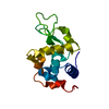
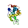
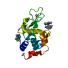
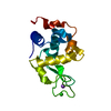
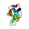
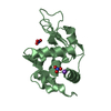
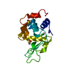
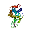
 Structure visualization
 Downloads & links
 Other downloads
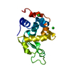


 PDBj
PDBj


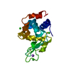
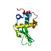
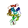
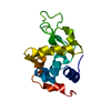
 Assembly


 Mass: 35.453 Da / Num. of mol.: 3 / Source method: obtained synthetically / Formula: Cl
 Mass: 22.990 Da / Num. of mol.: 1 / Source method: obtained synthetically / Formula: Na
 Mass: 18.015 Da / Num. of mol.: 89 / Source method: isolated from a natural source / Formula: H2O
 Sample preparation
 Processing