 Movie
Movie Controller

[English] 日本語
 Yorodumi
- PDB-4zi3: BART-like domain of BARTL1/CCDC104 aa1-133 in complex with Arl3FL... -
+ Open data

- Basic information
| Entry | Database: PDB / ID: 4zi3 | ||||||
|---|---|---|---|---|---|---|---|
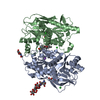
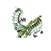
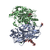
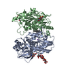
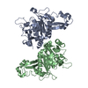
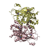
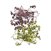
| Title | BART-like domain of BARTL1/CCDC104 aa1-133 in complex with Arl3FL bound to GppNHp in P1 21 1 | ||||||
 Components Components |
| ||||||
 Keywords Keywords | HYDROLASE / Arf-like GTPase / GTP-binding / BART-like domain / cilia | ||||||
| Function / homology |  Function and homology information Function and homology informationTrafficking of myristoylated proteins to the cilium / photoreceptor cell development / protein localization to ciliary membrane / intraciliary transport / post-Golgi vesicle-mediated transport / photoreceptor connecting cilium / Golgi to plasma membrane transport / protein localization to cilium / motile cilium / smoothened signaling pathway ...Trafficking of myristoylated proteins to the cilium / photoreceptor cell development / protein localization to ciliary membrane / intraciliary transport / post-Golgi vesicle-mediated transport / photoreceptor connecting cilium / Golgi to plasma membrane transport / protein localization to cilium / motile cilium / smoothened signaling pathway / small GTPase-mediated signal transduction / ciliary base / ciliary transition zone / mitotic cytokinesis / cilium assembly / axoneme / cytoplasmic microtubule / spindle microtubule / kidney development / GDP binding / protein transport / microtubule cytoskeleton / midbody / microtubule binding / cilium / ciliary basal body / Golgi membrane / GTPase activity / centrosome / GTP binding / magnesium ion binding / Golgi apparatus / nucleoplasm / nucleus / cytoplasm Similarity search - Function | ||||||
| Biological species |  | ||||||
| Method |  X-RAY DIFFRACTION / SYNCHROTRON / MOLECULAR REPLACEMENT / Resolution: 2 Å X-RAY DIFFRACTION / SYNCHROTRON / MOLECULAR REPLACEMENT / Resolution: 2 Å | ||||||
 Authors Authors | Lokaj, M. / Koerner, C. / Koesling, S. / Wittinghofer, A. | ||||||
| Funding support |  Germany, 1items Germany, 1items
| ||||||
 Citation Citation | Journal: Structure / Year: 2015 Title: The Interaction of CCDC104/BARTL1 with Arl3 and Implications for Ciliary Function. Authors: Lokaj, M. / Kosling, S.K. / Koerner, C. / Lange, S.M. / van Beersum, S.E. / van Reeuwijk, J. / Roepman, R. / Horn, N. / Ueffing, M. / Boldt, K. / Wittinghofer, A. | ||||||
| History |
|
- Structure visualization
| Structure viewer | Molecule: MolmilJmol/JSmol |
|---|
- Downloads & links
-Download
| PDBx/mmCIF format | 4zi3.cif.gz | 148.1 KB | Display | PDBx/mmCIF format |
|---|---|---|---|---|
| PDB format | pdb4zi3.ent.gz | 114.8 KB | Display | PDB format |
| PDBx/mmJSON format | 4zi3.json.gz | Tree view | PDBx/mmJSON format | |
| Others |  Other downloads Other downloads |
-Validation report
| Arichive directory | https://data.pdbj.org/pub/pdb/validation_reports/zi/4zi3ftp://data.pdbj.org/pub/pdb/validation_reports/zi/4zi3 | HTTPS FTP |
|---|
-Related structure data
| Related structure data | 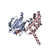 4zi2SC S: Starting model for refinement C: citing same article ( |
|---|---|
| Similar structure data |

-Links
 PDBj
PDBj


- Assembly
| Deposited unit | 
| ||||||||
|---|---|---|---|---|---|---|---|---|---|
| 1 |
| ||||||||
| Unit cell |
|
-Components
-Protein , 1 types, 2 molecules AB
| #1: Protein | Mass: 21582.654 Da / Num. of mol.: 2 Source method: isolated from a genetically manipulated source Source: (gene. exp.)  |
|---|
-Cilia- and flagella-associated protein ... , 2 types, 2 molecules CD
| #2: Protein | Mass: 15570.520 Da / Num. of mol.: 1 / Fragment: UNP residues 1-133 Source method: isolated from a genetically manipulated source Source: (gene. exp.) |
|---|---|
| #3: Protein | Mass: 15416.351 Da / Num. of mol.: 1 / Fragment: UNP residues 1-133 Source method: isolated from a genetically manipulated source Source: (gene. exp.) |
-Non-polymers , 3 types, 278 molecules 


| #4: Chemical |  Mass: 522.196 Da / Num. of mol.: 2 / Source method: obtained synthetically / Formula: C10H17N6O13P3 Mass: 522.196 Da / Num. of mol.: 2 / Source method: obtained synthetically / Formula: C10H17N6O13P3Comment: GppNHp, GMPPNP, energy-carrying molecule analogue*YM #5: Chemical |  Mass: 24.305 Da / Num. of mol.: 2 / Source method: obtained synthetically / Formula: Mg Mass: 24.305 Da / Num. of mol.: 2 / Source method: obtained synthetically / Formula: Mg#6: Water | ChemComp-HOH / | Mass: 18.015 Da / Num. of mol.: 274 / Source method: isolated from a natural source / Formula: H2O |
|---|
-Experimental details
-Experiment
| Experiment | Method: X-RAY DIFFRACTION |
|---|
- Sample preparation
| Crystal | Density Matthews: 2.26 Å3/Da / Density % sol: 45.61 % |
|---|---|
| Crystal grow | Temperature: 293.15 K / Method: vapor diffusion, hanging drop / pH: 8.5 / Details: 1 M LiCl, 0.1 M Tris pH8.5, 20 % PEG 4000 |
-Data collection
| Diffraction | Mean temperature: 100 K |
|---|---|
| Diffraction source | Source: SYNCHROTRON / Site: SLS  / Beamline: X10SA / Wavelength: 1 Å / Beamline: X10SA / Wavelength: 1 Å |
| Detector | Type: PSI PILATUS 6M / Detector: PIXEL / Date: Sep 6, 2013 |
| Radiation | Protocol: SINGLE WAVELENGTH / Monochromatic (M) / Laue (L): M / Scattering type: x-ray |
| Radiation wavelength | Wavelength: 1 Å / Relative weight: 1 |
| Reflection | Resolution: 2→28.95 Å / Num. all: 51617 / Num. obs: 44692 / % possible obs: 99.2 % / Redundancy: 3.35 % / Rmerge(I) obs: 0.058 / Net I/σ(I): 15.28 |
| Reflection shell | Resolution: 2→2.1 Å / Redundancy: 3.24 % / Mean I/σ(I) obs: 3.72 / % possible all: 99.1 |
- Processing
| Software |
| |||||||||||||||||||||||||||||||||||||||||||||||||||||||||||||||||||||||||||||||||||||||||||||||||||||||||||||||||||||||
|---|---|---|---|---|---|---|---|---|---|---|---|---|---|---|---|---|---|---|---|---|---|---|---|---|---|---|---|---|---|---|---|---|---|---|---|---|---|---|---|---|---|---|---|---|---|---|---|---|---|---|---|---|---|---|---|---|---|---|---|---|---|---|---|---|---|---|---|---|---|---|---|---|---|---|---|---|---|---|---|---|---|---|---|---|---|---|---|---|---|---|---|---|---|---|---|---|---|---|---|---|---|---|---|---|---|---|---|---|---|---|---|---|---|---|---|---|---|---|---|---|
| Refinement | Method to determine structure: MOLECULAR REPLACEMENT Starting model: 4ZI2 Resolution: 2→28.95 Å / SU ML: 0.25 / Cross valid method: FREE R-VALUE / σ(F): 1.37 / Phase error: 25.59 / Stereochemistry target values: ML
| |||||||||||||||||||||||||||||||||||||||||||||||||||||||||||||||||||||||||||||||||||||||||||||||||||||||||||||||||||||||
| Solvent computation | Shrinkage radii: 0.9 Å / VDW probe radii: 1.11 Å / Solvent model: FLAT BULK SOLVENT MODEL | |||||||||||||||||||||||||||||||||||||||||||||||||||||||||||||||||||||||||||||||||||||||||||||||||||||||||||||||||||||||
| Refinement step | Cycle: LAST / Resolution: 2→28.95 Å
| |||||||||||||||||||||||||||||||||||||||||||||||||||||||||||||||||||||||||||||||||||||||||||||||||||||||||||||||||||||||
| Refine LS restraints |
| |||||||||||||||||||||||||||||||||||||||||||||||||||||||||||||||||||||||||||||||||||||||||||||||||||||||||||||||||||||||
| LS refinement shell |
|
