 Movie
Movie Controller


+ Open data

- Basic information
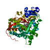
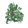
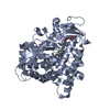
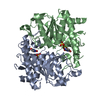
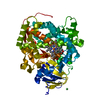
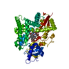
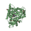
| Entry | Database: PDB / ID: 4zf6 | ||||||
|---|---|---|---|---|---|---|---|
| Title | Cytochrome P450 pentamutant from BM3 with bound PEG | ||||||
 Components Components | Bifunctional P-450/NADPH-P450 reductase | ||||||
 Keywords Keywords | OXIDOREDUCTASE / Cytochrome P450 / Heme Oxidase Domain / Bacillus megaterium | ||||||
| Function / homology |  Function and homology information Function and homology informationaromatase activity / NADPH-hemoprotein reductase / NADPH-hemoprotein reductase activity / oxidoreductase activity, acting on paired donors, with incorporation or reduction of molecular oxygen, reduced flavin or flavoprotein as one donor, and incorporation of one atom of oxygen / unspecific monooxygenase / FMN binding / flavin adenine dinucleotide binding / iron ion binding / heme binding / identical protein binding / cytosol Similarity search - Function | ||||||
| Biological species |  Bacillus megaterium (bacteria) Bacillus megaterium (bacteria) | ||||||
| Method |  X-RAY DIFFRACTION / SYNCHROTRON / MOLECULAR REPLACEMENT / Resolution: 2.773 Å X-RAY DIFFRACTION / SYNCHROTRON / MOLECULAR REPLACEMENT / Resolution: 2.773 Å | ||||||
 Authors Authors | Rogers, W.E. / Othman, T. / Heidary, D.K. / Huxford, T. | ||||||
 Citation Citation | Journal: Biochemistry / Year: 2016 Title: Effect of Mutation and Substrate Binding on the Stability of Cytochrome P450BM3 Variants. Authors: Geronimo, I. / Denning, C.A. / Rogers, W.E. / Othman, T. / Huxford, T. / Heidary, D.K. / Glazer, E.C. / Payne, C.M. | ||||||
| History |
|
- Structure visualization
| Structure viewer | Molecule: MolmilJmol/JSmol |
|---|
- Downloads & links
-Download
| PDBx/mmCIF format | 4zf6.cif.gz | 111.1 KB | Display | PDBx/mmCIF format |
|---|---|---|---|---|
| PDB format | pdb4zf6.ent.gz | 83.1 KB | Display | PDB format |
| PDBx/mmJSON format | 4zf6.json.gz | Tree view | PDBx/mmJSON format | |
| Others |  Other downloads Other downloads |
-Validation report
| Arichive directory | https://data.pdbj.org/pub/pdb/validation_reports/zf/4zf6ftp://data.pdbj.org/pub/pdb/validation_reports/zf/4zf6 | HTTPS FTP |
|---|
-Related structure data
| Related structure data |  4zf8C  4zfaC  4zfbC  3nplS  4zf9 4zfd 4zfe C: citing same article ( S: Starting model for refinement |
|---|---|
| Similar structure data |


-Links
 PDBj
PDBj


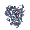
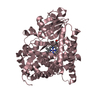
- Assembly
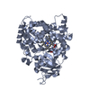
| Deposited unit | 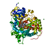
| ||||||||
|---|---|---|---|---|---|---|---|---|---|
| 1 |
| ||||||||
| Unit cell |
|
-Components
-Protein , 1 types, 1 molecules A
| #1: Protein | Mass: 52571.000 Da / Num. of mol.: 1 / Fragment: UNP residues 1-461 / Mutation: R47L, F81I, F87V, L188Q, E267V Source method: isolated from a genetically manipulated source Source: (gene. exp.) Bacillus megaterium (bacteria) / Gene: cyp102A1, cyp102 / Production host: References: UniProt: P14779, unspecific monooxygenase, NADPH-hemoprotein reductase |
|---|
-Non-polymers , 5 types, 66 molecules 


| #2: Chemical | ChemComp-HEM /  Mass: 616.487 Da / Num. of mol.: 1 / Source method: isolated from a natural source / Formula: C34H32FeN4O4 Mass: 616.487 Da / Num. of mol.: 1 / Source method: isolated from a natural source / Formula: C34H32FeN4O4 | ||
|---|---|---|---|
| #3: Chemical | ChemComp-1PE /  Mass: 238.278 Da / Num. of mol.: 1 / Source method: obtained synthetically / Formula: C10H22O6 / Comment: precipitant*YM Mass: 238.278 Da / Num. of mol.: 1 / Source method: obtained synthetically / Formula: C10H22O6 / Comment: precipitant*YM | ||
| #4: Chemical | ChemComp-EDO /  Mass: 62.068 Da / Num. of mol.: 1 / Source method: obtained synthetically / Formula: C2H6O2 Mass: 62.068 Da / Num. of mol.: 1 / Source method: obtained synthetically / Formula: C2H6O2 | ||
| #5: Chemical | ChemComp-NI /  Mass: 58.693 Da / Num. of mol.: 4 / Source method: obtained synthetically / Formula: Ni Mass: 58.693 Da / Num. of mol.: 4 / Source method: obtained synthetically / Formula: Ni#6: Water | ChemComp-HOH / | Mass: 18.015 Da / Num. of mol.: 59 / Source method: isolated from a natural source / Formula: H2O |
-Experimental details
-Experiment
| Experiment | Method: X-RAY DIFFRACTION / Number of used crystals: 1 |
|---|
- Sample preparation
| Crystal | Density Matthews: 3.07 Å3/Da / Density % sol: 59.9 % |
|---|---|
| Crystal grow | Temperature: 298 K / Method: vapor diffusion, hanging drop / Details: 17.5mM NiCl2, 50mM PEG MME 2000 |
-Data collection
| Diffraction | Mean temperature: 100 K |
|---|---|
| Diffraction source | Source: SYNCHROTRON / Site: APS  / Beamline: 24-ID-C / Wavelength: 0.9792 Å / Beamline: 24-ID-C / Wavelength: 0.9792 Å |
| Detector | Type: DECTRIS PILATUS 6M-F / Detector: PIXEL / Date: Oct 31, 2013 |
| Radiation | Protocol: SINGLE WAVELENGTH / Monochromatic (M) / Laue (L): M / Scattering type: x-ray |
| Radiation wavelength | Wavelength: 0.9792 Å / Relative weight: 1 |
| Reflection | Resolution: 2.77→177.81 Å / Num. obs: 18183 / % possible obs: 97.2 % / Redundancy: 7.5 % / Rmerge(I) obs: 0.07 / Net I/σ(I): 17.7 |
| Reflection shell | Resolution: 2.77→2.92 Å / Redundancy: 3.4 % / Rmerge(I) obs: 0.323 / Mean I/σ(I) obs: 3.4 / % possible all: 80.6 |
- Processing
| Software |
| ||||||||||||||||||||||||||||||||||||||||||||||||||||||||
|---|---|---|---|---|---|---|---|---|---|---|---|---|---|---|---|---|---|---|---|---|---|---|---|---|---|---|---|---|---|---|---|---|---|---|---|---|---|---|---|---|---|---|---|---|---|---|---|---|---|---|---|---|---|---|---|---|---|
| Refinement | Method to determine structure: MOLECULAR REPLACEMENT Starting model: 3NPL Resolution: 2.773→59.269 Å / SU ML: 0.4 / Cross valid method: FREE R-VALUE / σ(F): 1.34 / Phase error: 24.13 / Stereochemistry target values: ML
| ||||||||||||||||||||||||||||||||||||||||||||||||||||||||
| Solvent computation | Shrinkage radii: 0.9 Å / VDW probe radii: 1.11 Å / Solvent model: FLAT BULK SOLVENT MODEL | ||||||||||||||||||||||||||||||||||||||||||||||||||||||||
| Refinement step | Cycle: LAST / Resolution: 2.773→59.269 Å
| ||||||||||||||||||||||||||||||||||||||||||||||||||||||||
| Refine LS restraints |
| ||||||||||||||||||||||||||||||||||||||||||||||||||||||||
| LS refinement shell |
|
