 Movie
Movie Controller

[English] 日本語
 Yorodumi
- PDB-4zda: Crystal structure of isocitrate dehydrogenase in complex with iso... -
+ Open data

- Basic information
| Entry | Database: PDB / ID: 4zda | ||||||
|---|---|---|---|---|---|---|---|
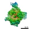
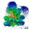
| Title | Crystal structure of isocitrate dehydrogenase in complex with isocitrate and Mn from M. smegmatis | ||||||
 Components Components | Isocitrate dehydrogenase (NADP) Icd2 | ||||||
 Keywords Keywords | OXIDOREDUCTASE / ICD / M. smegmatis / complex / Isocitrate | ||||||
| Function / homology |  Function and homology information Function and homology informationisocitrate dehydrogenase (NADP+) / isocitrate dehydrogenase (NADP+) activity / glyoxylate cycle / tricarboxylic acid cycle / metal ion binding Similarity search - Function | ||||||
| Biological species |  Mycobacterium smegmatis (bacteria) Mycobacterium smegmatis (bacteria) | ||||||
| Method |  X-RAY DIFFRACTION / SYNCHROTRON / MOLECULAR REPLACEMENT / Resolution: 2.8 Å X-RAY DIFFRACTION / SYNCHROTRON / MOLECULAR REPLACEMENT / Resolution: 2.8 Å | ||||||
 Authors Authors | Pojer, F. / Murima, P. / McKinney, J.D. | ||||||
 Citation Citation | Journal: To Be Published Title: crystal structure of isocitrate dehydrogenase in complex with isocitrate and Mn from M. smegmatis Authors: Pojer, F. / Murima, P. / McKinney, J.D. | ||||||
| History |
|
- Structure visualization
| Structure viewer | Molecule: MolmilJmol/JSmol |
|---|
- Downloads & links
-Download
| PDBx/mmCIF format | 4zda.cif.gz | 823.1 KB | Display | PDBx/mmCIF format |
|---|---|---|---|---|
| PDB format | pdb4zda.ent.gz | 687.8 KB | Display | PDB format |
| PDBx/mmJSON format | 4zda.json.gz | Tree view | PDBx/mmJSON format | |
| Others |  Other downloads Other downloads |
-Validation report
| Arichive directory | https://data.pdbj.org/pub/pdb/validation_reports/zd/4zdaftp://data.pdbj.org/pub/pdb/validation_reports/zd/4zda | HTTPS FTP |
|---|
-Related structure data
| Similar structure data |
|---|
-Links
 PDBj
PDBj


- Assembly
| Deposited unit | 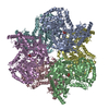
| ||||||||
|---|---|---|---|---|---|---|---|---|---|
| 1 |
| ||||||||
| Unit cell |
|
-Components
| #1: Protein | Mass: 85581.336 Da / Num. of mol.: 6 Source method: isolated from a genetically manipulated source Source: (gene. exp.) Mycobacterium smegmatis (bacteria) / Gene: icd2, MSMEG_1654, MSMEI_1615 / Production host: References: UniProt: A0QSZ3, isocitrate dehydrogenase (NADP+) #2: Chemical | ChemComp-MN /   Mass: 54.938 Da / Num. of mol.: 6 / Source method: obtained synthetically / Formula: Mn Mass: 54.938 Da / Num. of mol.: 6 / Source method: obtained synthetically / Formula: Mn#3: Chemical | ChemComp-ICT /   Mass: 192.124 Da / Num. of mol.: 6 / Source method: obtained synthetically / Formula: C6H8O7 Mass: 192.124 Da / Num. of mol.: 6 / Source method: obtained synthetically / Formula: C6H8O7 |
|---|
-Experimental details
-Experiment
| Experiment | Method: X-RAY DIFFRACTION |
|---|
- Sample preparation
| Crystal | Density Matthews: 2.95 Å3/Da / Density % sol: 58.28 % |
|---|---|
| Crystal grow | Temperature: 291 K / Method: vapor diffusion, hanging drop Details: 0.05 M MgCl2, 28-34% Polyethylene glycol monomethyl ether 550 (Qiagen), and 0.1M Hepes pH 7.5 |
-Data collection
| Diffraction | Mean temperature: 100 K |
|---|---|
| Diffraction source | Source: SYNCHROTRON / Site: SLS  / Beamline: X06DA / Wavelength: 1 Å / Beamline: X06DA / Wavelength: 1 Å |
| Detector | Type: PSI PILATUS 6M / Detector: PIXEL / Date: Jun 24, 2013 |
| Radiation | Protocol: SINGLE WAVELENGTH / Monochromatic (M) / Laue (L): M / Scattering type: x-ray |
| Radiation wavelength | Wavelength: 1 Å / Relative weight: 1 |
| Reflection | Resolution: 2.8→50 Å / Num. obs: 145405 / % possible obs: 97.4 % / Redundancy: 2.16 % / Net I/σ(I): 12.81 |
- Processing
| Software |
| ||||||||||||||||||||||||||||||||||||||||||||||||||||||||||||||||||||||||||||||||||||||||||||||||||||||||||||||||||||||||||||||||||||||||||||||||||||||||||||||||||||||||||||||||||||||
|---|---|---|---|---|---|---|---|---|---|---|---|---|---|---|---|---|---|---|---|---|---|---|---|---|---|---|---|---|---|---|---|---|---|---|---|---|---|---|---|---|---|---|---|---|---|---|---|---|---|---|---|---|---|---|---|---|---|---|---|---|---|---|---|---|---|---|---|---|---|---|---|---|---|---|---|---|---|---|---|---|---|---|---|---|---|---|---|---|---|---|---|---|---|---|---|---|---|---|---|---|---|---|---|---|---|---|---|---|---|---|---|---|---|---|---|---|---|---|---|---|---|---|---|---|---|---|---|---|---|---|---|---|---|---|---|---|---|---|---|---|---|---|---|---|---|---|---|---|---|---|---|---|---|---|---|---|---|---|---|---|---|---|---|---|---|---|---|---|---|---|---|---|---|---|---|---|---|---|---|---|---|---|---|
| Refinement | Method to determine structure: MOLECULAR REPLACEMENT / Resolution: 2.8→48.59 Å / Cor.coef. Fo:Fc: 0.941 / Cor.coef. Fo:Fc free: 0.914 / SU B: 15.905 / SU ML: 0.304 / Cross valid method: THROUGHOUT / ESU R Free: 0.367 / Stereochemistry target values: MAXIMUM LIKELIHOOD / Details: HYDROGENS HAVE BEEN USED IF PRESENT IN THE INPUT
| ||||||||||||||||||||||||||||||||||||||||||||||||||||||||||||||||||||||||||||||||||||||||||||||||||||||||||||||||||||||||||||||||||||||||||||||||||||||||||||||||||||||||||||||||||||||
| Solvent computation | Ion probe radii: 0.8 Å / Shrinkage radii: 0.8 Å / VDW probe radii: 1.2 Å / Solvent model: MASK | ||||||||||||||||||||||||||||||||||||||||||||||||||||||||||||||||||||||||||||||||||||||||||||||||||||||||||||||||||||||||||||||||||||||||||||||||||||||||||||||||||||||||||||||||||||||
| Displacement parameters | Biso mean: 68.633 Å2
| ||||||||||||||||||||||||||||||||||||||||||||||||||||||||||||||||||||||||||||||||||||||||||||||||||||||||||||||||||||||||||||||||||||||||||||||||||||||||||||||||||||||||||||||||||||||
| Refinement step | Cycle: 1 / Resolution: 2.8→48.59 Å
| ||||||||||||||||||||||||||||||||||||||||||||||||||||||||||||||||||||||||||||||||||||||||||||||||||||||||||||||||||||||||||||||||||||||||||||||||||||||||||||||||||||||||||||||||||||||
| Refine LS restraints |
|