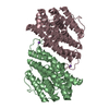
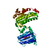
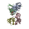
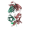
[English] 日本語
Yorodumi
- PDB-4zbw: Crystal structure of death effector domain of Caspase8 in Homo Sapiens -

+
Open data


ID or keywords:

Loading...

-
Basic information

Entry
Database: PDB / ID: 4zbw
TitleCrystal structure of death effector domain of Caspase8 in Homo Sapiens
ComponentsCaspase-8
KeywordsHYDROLASE / Death effector domain / caspase
Function / homology
Function and homology information


caspase-8 / death effector domain binding / FasL/ CD95L signaling / syncytiotrophoblast cell differentiation involved in labyrinthine layer development / TRAIL signaling / CD95 death-inducing signaling complex / ripoptosome / Defective RIPK1-mediated regulated necrosis / TRAIL-activated apoptotic signaling pathway / Apoptotic execution phase ...caspase-8 / death effector domain binding / FasL/ CD95L signaling / syncytiotrophoblast cell differentiation involved in labyrinthine layer development / TRAIL signaling / CD95 death-inducing signaling complex / ripoptosome / Defective RIPK1-mediated regulated necrosis / TRAIL-activated apoptotic signaling pathway / Apoptotic execution phase / Activation, myristolyation of BID and translocation to mitochondria / TRIF-mediated programmed cell death / Microbial modulation of RIPK1-mediated regulated necrosis / Regulation by c-FLIP / CASP8 activity is inhibited / Dimerization of procaspase-8 / TLR3-mediated TICAM1-dependent programmed cell death / Caspase activation via Death Receptors in the presence of ligand / positive regulation of macrophage differentiation / self proteolysis / response to cobalt ion / NF-kB activation through FADD/RIP-1 pathway mediated by caspase-8 and -10 / death-inducing signaling complex / CLEC7A/inflammasome pathway / negative regulation of necroptotic process / response to anesthetic / regulation of tumor necrosis factor-mediated signaling pathway / tumor necrosis factor receptor binding / death receptor binding / natural killer cell activation / TNFR1-induced proapoptotic signaling / RIPK1-mediated regulated necrosis / execution phase of apoptosis / regulation of innate immune response / Apoptotic cleavage of cellular proteins / pyroptotic inflammatory response / : / B cell activation / positive regulation of proteolysis / macrophage differentiation / response to tumor necrosis factor / extrinsic apoptotic signaling pathway via death domain receptors / Caspase-mediated cleavage of cytoskeletal proteins / negative regulation of canonical NF-kappaB signal transduction / extrinsic apoptotic signaling pathway / cysteine-type peptidase activity / regulation of cytokine production / protein maturation / proteolysis involved in protein catabolic process / T cell activation / positive regulation of interleukin-1 beta production / Regulation of NF-kappa B signaling / apoptotic signaling pathway / Regulation of TNFR1 signaling / NOD1/2 Signaling Pathway / protein processing / Regulation of necroptotic cell death / cellular response to mechanical stimulus / positive regulation of neuron apoptotic process / response to estradiol / lamellipodium / peptidase activity / heart development / cell body / scaffold protein binding / angiogenesis / response to lipopolysaccharide / response to ethanol / mitochondrial outer membrane / positive regulation of canonical NF-kappaB signal transduction / cytoskeleton / positive regulation of cell migration / positive regulation of apoptotic process / cysteine-type endopeptidase activity / apoptotic process / ubiquitin protein ligase binding / protein-containing complex binding / protein-containing complex / mitochondrion / proteolysis / identical protein binding / nucleus / cytosol / cytoplasm
Similarity search - Function
Caspase-8 / Death effector domain / Death effector domain (DED) profile. / Death effector domain / Death effector domain / Death Domain, Fas / Death Domain, Fas / Peptidase family C14A, His active site / Caspase family histidine active site. / Peptidase C14, caspase non-catalytic subunit p10 ...Caspase-8 / Death effector domain / Death effector domain (DED) profile. / Death effector domain / Death effector domain / Death Domain, Fas / Death Domain, Fas / Peptidase family C14A, His active site / Caspase family histidine active site. / Peptidase C14, caspase non-catalytic subunit p10 / Peptidase family C14A, cysteine active site / Caspase family cysteine active site. / Caspase family p10 domain profile. / Peptidase C14A, caspase catalytic domain / Caspase, interleukin-1 beta converting enzyme (ICE) homologues / Peptidase C14, p20 domain / Caspase family p20 domain profile. / : / Caspase domain / Caspase-like domain superfamily / Death-like domain superfamily / Orthogonal Bundle / Mainly Alpha
Similarity search - Domain/homology
Biological speciesHomo sapiens (human)
MethodX-RAY DIFFRACTION / MOLECULAR REPLACEMENT / Resolution: 2.2 Å
AuthorsShen, C. / Wang, T. / Quan, J.
Funding support China, 3items
OrganizationGrant numberCountry
MOST2012CB722602 China
MOST2013CB911501 China
SZSTIGGJS20130329180714793 China
CitationJournal: Biochem.Biophys.Res.Commun. / Year: 2015
Title: Crystal structure of the death effector domains of caspase-8
Authors: Shen, C. / Yue, H. / Pei, J. / Guo, X. / Wang, T. / Quan, J.M.
History
DepositionApr 15, 2015Deposition site: RCSB / Processing site: PDBJ
Revision 1.0Jul 1, 2015Provider: repository / Type: Initial release
Revision 1.1Nov 8, 2023Group: Data collection / Database references ...Data collection / Database references / Derived calculations / Refinement description / Source and taxonomy / Structure summary
Category: chem_comp_atom / chem_comp_bond ...chem_comp_atom / chem_comp_bond / citation / database_2 / entity_src_gen / pdbx_initial_refinement_model / pdbx_struct_assembly / pdbx_struct_assembly_gen / pdbx_struct_assembly_prop / pdbx_struct_oper_list / struct_keywords
Item: _citation.journal_id_CSD / _database_2.pdbx_DOI ..._citation.journal_id_CSD / _database_2.pdbx_DOI / _database_2.pdbx_database_accession / _entity_src_gen.pdbx_alt_source_flag / _pdbx_struct_assembly.oligomeric_details / _pdbx_struct_assembly_gen.asym_id_list / _pdbx_struct_assembly_prop.type / _pdbx_struct_assembly_prop.value / _pdbx_struct_oper_list.symmetry_operation / _struct_keywords.text

-
Structure visualization

Structure viewerMolecule:
MolmilJmol/JSmol

Downloads & links

-
Assembly

Deposited unit
A: Caspase-8
B: Caspase-8


Theoretical massNumber of molelcules
Total (without water)44,9782
Polymers44,9782
Non-polymers00
Water4,468248
1


  • Idetical with deposited unit
  • defined by author
TypeNameSymmetry operationNumber
identity operation1_555x,y,z1
Buried area910 Å2
ΔGint2 kcal/mol
Surface area20260 Å2
Unit cell
Length a, b, c (Å)51.360, 52.106, 56.596
Angle α, β, γ (deg.)113.34, 116.89, 90.12
Int Tables number1
Space group name H-MP1

-
Components

#1: Protein Caspase-8 / CASP-8 / Apoptotic cysteine protease / Apoptotic protease Mch-5 / CAP4 / FADD-homologous ICE/ced-3- ...CASP-8 / Apoptotic cysteine protease / Apoptotic protease Mch-5 / CAP4 / FADD-homologous ICE/ced-3-like protease / FADD-like ICE / FLICE / ICE-like apoptotic protease 5 / MORT1-associated ced-3 homolog / MACH


Mass: 22488.879 Da / Num. of mol.: 2 / Mutation: F122A, I128D
Source method: isolated from a genetically manipulated source
Source: (gene. exp.) Homo sapiens (human) / Gene: CASP8, MCH5 / Plasmid: pET28a(+) / Production host: Escherichia coli (E. coli) / Strain (production host): Rosetta DE3 plysS / References: UniProt: Q14790, caspase-8
#2: Water ChemComp-HOH / water


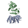
Mass: 18.015 Da / Num. of mol.: 248 / Source method: isolated from a natural source / Formula: H2O

-
Experimental details

-
Experiment

ExperimentMethod: X-RAY DIFFRACTION

-
Sample preparation

CrystalDensity Matthews: 2.6 Å3/Da / Density % sol: 54.26 % / Description: rod like crystal, 0.1X0.1X0.4
Crystal growTemperature: 293 K / Method: evaporation / pH: 8.5 / Details: 0.2M sodium chloride, 25% PEG3350 / PH range: 8.0-8.5

-
Data collection

DiffractionMean temperature: 100 K
Diffraction sourceSource: SEALED TUBE / Type: RIGAKU MICROMAX-002+ / Wavelength: 1.5418 Å
DetectorType: RIGAKU SATURN 944+ / Detector: CCD / Date: Jun 27, 2014
RadiationMonochromator: Confocal / Protocol: SINGLE WAVELENGTH / Monochromatic (M) / Laue (L): M / Scattering type: x-ray
Radiation wavelengthWavelength: 1.5418 Å / Relative weight: 1
ReflectionResolution: 2.2→29.22 Å / Num. obs: 22971 / % possible obs: 96.6 % / Redundancy: 1.83 % / Biso Wilson estimate: 30.98 Å2 / Rmerge(I) obs: 0.052 / Net I/σ(I): 9
Reflection shellResolution: 2.2→2.28 Å / Redundancy: 1.59 % / Rmerge(I) obs: 0.238 / Mean I/σ(I) obs: 2.6 / % possible all: 93.8

-
Processing

Software
NameVersionClassification
PHENIX1.9_1692refinement
CrystalClear1.4data reduction
d*TREK9.7data scaling
PHASER2.5.6phasing
RefinementMethod to determine structure: MOLECULAR REPLACEMENT
Starting model: 2BBR
Resolution: 2.2→29.22 Å / SU ML: 0.3 / Cross valid method: NONE / σ(F): 1.96 / Phase error: 28.05 / Stereochemistry target values: ML
RfactorNum. reflection% reflectionSelection details
Rfree0.251 2010 8.76 %Random
Rwork0.2041 ---
obs0.2082 22952 96.5 %-
Solvent computationShrinkage radii: 0.9 Å / VDW probe radii: 1.11 Å / Solvent model: FLAT BULK SOLVENT MODEL
Displacement parametersBiso mean: 41.3 Å2
Refinement stepCycle: LAST / Resolution: 2.2→29.22 Å
ProteinNucleic acidLigandSolventTotal
Num. atoms3050 0 0 248 3298
Refine LS restraints
Refine-IDTypeDev idealNumber
X-RAY DIFFRACTIONf_bond_d0.0043082
X-RAY DIFFRACTIONf_angle_d0.84120
X-RAY DIFFRACTIONf_dihedral_angle_d14.6831248
X-RAY DIFFRACTIONf_chiral_restr0.029462
X-RAY DIFFRACTIONf_plane_restr0.005532
LS refinement shell
Resolution (Å)Rfactor RfreeNum. reflection RfreeRfactor RworkNum. reflection RworkRefine-ID% reflection obs (%)
2.2-2.2550.34821440.28141417X-RAY DIFFRACTION92
2.255-2.3160.33521270.25561518X-RAY DIFFRACTION97
2.316-2.38410.28261610.24211500X-RAY DIFFRACTION98
2.3841-2.4610.30591400.23471544X-RAY DIFFRACTION98
2.461-2.54890.27161390.23361510X-RAY DIFFRACTION99
2.5489-2.65090.27461380.23211517X-RAY DIFFRACTION98
2.6509-2.77150.28891510.231545X-RAY DIFFRACTION98
2.7715-2.91740.30991380.24181528X-RAY DIFFRACTION98
2.9174-3.10.33521440.23441515X-RAY DIFFRACTION98
3.1-3.33910.26241440.2251550X-RAY DIFFRACTION97
3.3391-3.67450.24271580.20211461X-RAY DIFFRACTION97
3.6745-4.20490.23841520.16981469X-RAY DIFFRACTION96
4.2049-5.29260.17331480.14911459X-RAY DIFFRACTION94
5.2926-29.22470.18511260.17381409X-RAY DIFFRACTION90
Refinement TLS params.

Method: refined / Refine-ID: X-RAY DIFFRACTION

IDL112)L122)L132)L222)L232)L332)S11 (Å °)S12 (Å °)S13 (Å °)S21 (Å °)S22 (Å °)S23 (Å °)S31 (Å °)S32 (Å °)S33 (Å °)T112)T122)T132)T222)T232)T332)Origin x (Å)Origin y (Å)Origin z (Å)
11.71570.84731.43791.98371.20352.7121-0.057-0.00050.1561-0.0878-0.02490.0928-0.08560.02950.05920.24540.00970.04370.24190.01450.2876-19.535422.4626-38.9367
21.7739-0.6627-1.30491.76731.02672.0844-0.04340.0371-0.18290.1065-0.0390.120.1169-0.01980.07380.1959-0.0001-0.03820.18740.01760.2216.1586-3.2216-42.4737
Refinement TLS group
IDRefine-IDRefine TLS-IDSelection details
1X-RAY DIFFRACTION1chain 'A'
2X-RAY DIFFRACTION2chain 'B'

+
About Yorodumi

-
News

-
Feb 9, 2022. New format data for meta-information of EMDB entries

New format data for meta-information of EMDB entries

  • Version 3 of the EMDB header file is now the official format.
  • The previous official version 1.9 will be removed from the archive.

Related info.:EMDB header

External links:wwPDB to switch to version 3 of the EMDB data model

-
Aug 12, 2020. Covid-19 info

Covid-19 info

URL: https://pdbj.org/emnavi/covid19.php

New page: Covid-19 featured information page in EM Navigator.

Related info.:Covid-19 info / Mar 5, 2020. Novel coronavirus structure data

+
Mar 5, 2020. Novel coronavirus structure data

Novel coronavirus structure data

Related info.:Yorodumi Speices / Aug 12, 2020. Covid-19 info

External links:COVID-19 featured content - PDBj / Molecule of the Month (242):Coronavirus Proteases

+
Jan 31, 2019. EMDB accession codes are about to change! (news from PDBe EMDB page)

EMDB accession codes are about to change! (news from PDBe EMDB page)

  • The allocation of 4 digits for EMDB accession codes will soon come to an end. Whilst these codes will remain in use, new EMDB accession codes will include an additional digit and will expand incrementally as the available range of codes is exhausted. The current 4-digit format prefixed with “EMD-” (i.e. EMD-XXXX) will advance to a 5-digit format (i.e. EMD-XXXXX), and so on. It is currently estimated that the 4-digit codes will be depleted around Spring 2019, at which point the 5-digit format will come into force.
  • The EM Navigator/Yorodumi systems omit the EMD- prefix.

Related info.:Q: What is EMD? / ID/Accession-code notation in Yorodumi/EM Navigator

External links:EMDB Accession Codes are Changing Soon! / Contact to PDBj

+
Jul 12, 2017. Major update of PDB

Major update of PDB

  • wwPDB released updated PDB data conforming to the new PDBx/mmCIF dictionary.
  • This is a major update changing the version number from 4 to 5, and with Remediation, in which all the entries are updated.
  • In this update, many items about electron microscopy experimental information are reorganized (e.g. em_software).
  • Now, EM Navigator and Yorodumi are based on the updated data.

External links:wwPDB Remediation / Enriched Model Files Conforming to OneDep Data Standards Now Available in the PDB FTP Archive

-
Yorodumi

Thousand views of thousand structures

  • Yorodumi is a browser for structure data from EMDB, PDB, SASBDB, etc.
  • This page is also the successor to EM Navigator detail page, and also detail information page/front-end page for Omokage search.
  • The word "yorodu" (or yorozu) is an old Japanese word meaning "ten thousand". "mi" (miru) is to see.

Related info.:EMDB / PDB / SASBDB / Comparison of 3 databanks / Yorodumi Search / Aug 31, 2016. New EM Navigator & Yorodumi / Yorodumi Papers / Jmol/JSmol / Function and homology information / Changes in new EM Navigator and Yorodumi

Read more