 Movie
Movie Controller

[English] 日本語
 Yorodumi
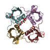
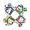
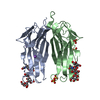
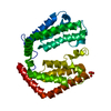
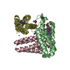
- PDB-4z9s: Non-covalent assembly of monoubiquitin that mimics K11 poly-ubiquitin -
+ Open data

- Basic information
| Entry | Database: PDB / ID: 4z9s | ||||||
|---|---|---|---|---|---|---|---|
| Title | Non-covalent assembly of monoubiquitin that mimics K11 poly-ubiquitin | ||||||
 Components Components | Ubiquitin-40S ribosomal protein S27a | ||||||
 Keywords Keywords | CELL CYCLE | ||||||
| Function / homology |  Function and homology information Function and homology informationTranslation initiation complex formation / Formation of the ternary complex, and subsequently, the 43S complex / Ribosomal scanning and start codon recognition / Translesion synthesis by REV1 / Recognition of DNA damage by PCNA-containing replication complex / Translesion Synthesis by POLH / Downregulation of ERBB4 signaling / Spry regulation of FGF signaling / Downregulation of ERBB2:ERBB3 signaling / NOD1/2 Signaling Pathway ...Translation initiation complex formation / Formation of the ternary complex, and subsequently, the 43S complex / Ribosomal scanning and start codon recognition / Translesion synthesis by REV1 / Recognition of DNA damage by PCNA-containing replication complex / Translesion Synthesis by POLH / Downregulation of ERBB4 signaling / Spry regulation of FGF signaling / Downregulation of ERBB2:ERBB3 signaling / NOD1/2 Signaling Pathway / APC/C:Cdc20 mediated degradation of Cyclin B / APC-Cdc20 mediated degradation of Nek2A / EGFR downregulation / TCF dependent signaling in response to WNT / NRIF signals cell death from the nucleus / p75NTR recruits signalling complexes / NF-kB is activated and signals survival / Activated NOTCH1 Transmits Signal to the Nucleus / Downregulation of TGF-beta receptor signaling / TGF-beta receptor signaling in EMT (epithelial to mesenchymal transition) / Downregulation of SMAD2/3:SMAD4 transcriptional activity / SMAD2/SMAD3:SMAD4 heterotrimer regulates transcription / Senescence-Associated Secretory Phenotype (SASP) / activated TAK1 mediates p38 MAPK activation / JNK (c-Jun kinases) phosphorylation and activation mediated by activated human TAK1 / Regulation of FZD by ubiquitination / N-glycan trimming in the ER and Calnexin/Calreticulin cycle / Regulation of TNFR1 signaling / TNFR1-induced NF-kappa-B signaling pathway / Translesion synthesis by POLK / Translesion synthesis by POLI / Regulation of necroptotic cell death / HDR through Homologous Recombination (HRR) / Josephin domain DUBs / Recruitment and ATM-mediated phosphorylation of repair and signaling proteins at DNA double strand breaks / Processing of DNA double-strand break ends / Gap-filling DNA repair synthesis and ligation in GG-NER / Dual Incision in GG-NER / Fanconi Anemia Pathway / Regulation of TP53 Activity through Phosphorylation / Regulation of TP53 Degradation / Regulation of TP53 Activity through Methylation / Negative regulation of MET activity / PTK6 Regulates RTKs and Their Effectors AKT1 and DOK1 / Downregulation of ERBB2 signaling / E3 ubiquitin ligases ubiquitinate target proteins / Regulation of PTEN localization / ER Quality Control Compartment (ERQC) / Regulation of expression of SLITs and ROBOs / Interferon alpha/beta signaling / Endosomal Sorting Complex Required For Transport (ESCRT) / Activation of IRF3, IRF7 mediated by TBK1, IKKε (IKBKE) / IKK complex recruitment mediated by RIP1 / IRAK2 mediated activation of TAK1 complex / TRAF6-mediated induction of TAK1 complex within TLR4 complex / Alpha-protein kinase 1 signaling pathway / RAS processing / Pexophagy / Negative regulation of FLT3 / IRAK2 mediated activation of TAK1 complex upon TLR7/8 or 9 stimulation / Regulation of NF-kappa B signaling / Regulation of TBK1, IKKε (IKBKE)-mediated activation of IRF3, IRF7 / Regulation of pyruvate metabolism / SCF-beta-TrCP mediated degradation of Emi1 / MAP3K8 (TPL2)-dependent MAPK1/3 activation / Ovarian tumor domain proteases / Cyclin D associated events in G1 / Regulation of BACH1 activity / Regulation of innate immune responses to cytosolic DNA / Negative regulation of FGFR1 signaling / Negative regulation of FGFR2 signaling / Negative regulation of FGFR3 signaling / Negative regulation of FGFR4 signaling / Negative regulation of MAPK pathway / Formation of Incision Complex in GG-NER / Synthesis of active ubiquitin: roles of E1 and E2 enzymes / Inactivation of CSF3 (G-CSF) signaling / Termination of translesion DNA synthesis / Iron uptake and transport / Negative regulators of DDX58/IFIH1 signaling / Deactivation of the beta-catenin transactivating complex / Metalloprotease DUBs / Formation of TC-NER Pre-Incision Complex / Dual incision in TC-NER / Gap-filling DNA repair synthesis and ligation in TC-NER / Major pathway of rRNA processing in the nucleolus and cytosol / GTP hydrolysis and joining of the 60S ribosomal subunit / Autodegradation of Cdh1 by Cdh1:APC/C / APC/C:Cdc20 mediated degradation of Securin / APC/C:Cdh1 mediated degradation of Cdc20 and other APC/C:Cdh1 targeted proteins in late mitosis/early G1 / Cdc20:Phospho-APC/C mediated degradation of Cyclin A / Autodegradation of the E3 ubiquitin ligase COP1 / Asymmetric localization of PCP proteins / Degradation of AXIN / Degradation of DVL / Hedgehog ligand biogenesis / Hedgehog 'on' state / TNFR2 non-canonical NF-kB pathway / DNA Damage Recognition in GG-NER / Assembly of the pre-replicative complex Similarity search - Function | ||||||
| Biological species |  | ||||||
| Method |  X-RAY DIFFRACTION / MOLECULAR REPLACEMENT / Resolution: 2.3 Å X-RAY DIFFRACTION / MOLECULAR REPLACEMENT / Resolution: 2.3 Å | ||||||
 Authors Authors | Levin-Kravets, O. / Prag, G. | ||||||
| Funding support | 1items
| ||||||
 Citation Citation | Journal: Biochemistry / Year: 2015 Title: Tetrameric Assembly of Monoubiquitin Accurately Mimics the Lys11 Polyubiquitin Chain Structure. Authors: Levin-Kravets, O. / Shohat, N. / Prag, G. #1: Journal: Nat.Struct.Mol.Biol. / Year: 2010Title: Lys11-Linked Ubiqutin Chains Adopt Compact Conformations And Are By The Deubiqutinase Cazannepreferentially Hydrolyzed Authors: Bremm, A. / Freund, S.M.V. / Komander, D. #2: Journal: J Mol Biol / Year: 1987Title: Structure of ubiquitin refined at 1.8 A resolution. Abstract: The crystal structure of human erythrocytic ubiquitin has been refined at 1.8 A resolution using a restrained least-squares procedure. The crystallographic R-factor for the final model is 0.176. Bond ...The crystal structure of human erythrocytic ubiquitin has been refined at 1.8 A resolution using a restrained least-squares procedure. The crystallographic R-factor for the final model is 0.176. Bond lengths and bond angles in the molecule have root-mean-square deviations from ideal values of 0.016 A and 1.5 degrees, respectively. A total of 58 water molecules per molecule of ubiquitin are included in the final model. The last four residues in the molecule appear to have partial occupancy or large thermal motion. The overall structure of ubiquitin is extremely compact and tightly hydrogen-bonded; approximately 87% of the polypeptide chain is involved in hydrogen-bonded secondary structure. Prominent secondary structural features include three and one-half turns of alpha-helix, a short piece of 3(10)-helix, a mixed beta-sheet that contains five strands, and seven reverse turns. There is a marked hydrophobic core formed between the beta-sheet and alpha-helix. The molecule features a number of unusual secondary structural features, including a parallel G1 beta-bulge, two reverse Asx turns, and a symmetrical hydrogen-bonding region that involves the two helices and two of the reverse turns. | ||||||
| History |
|
- Structure visualization
| Structure viewer | Molecule: MolmilJmol/JSmol |
|---|
- Downloads & links
-Download
| PDBx/mmCIF format | 4z9s.cif.gz | 83 KB | Display | PDBx/mmCIF format |
|---|---|---|---|---|
| PDB format | pdb4z9s.ent.gz | 64.7 KB | Display | PDB format |
| PDBx/mmJSON format | 4z9s.json.gz | Tree view | PDBx/mmJSON format | |
| Others |  Other downloads Other downloads |
-Validation report
| Arichive directory | https://data.pdbj.org/pub/pdb/validation_reports/z9/4z9sftp://data.pdbj.org/pub/pdb/validation_reports/z9/4z9s | HTTPS FTP |
|---|
-Related structure data
| Related structure data |  1ubqS S: Starting model for refinement |
|---|---|
| Similar structure data |


-Links
 PDBj
PDBj


- Assembly
| Deposited unit | 
| ||||||||||||
|---|---|---|---|---|---|---|---|---|---|---|---|---|---|
| 1 |
| ||||||||||||
| Unit cell |
| ||||||||||||
| Components on special symmetry positions |
|
-Components
| #1: Protein | Mass: 8576.831 Da / Num. of mol.: 4 / Fragment: UNP RESIDUES 1-76 Source method: isolated from a genetically manipulated source Source: (gene. exp.)  #2: Chemical | ChemComp-MLA /   Mass: 104.061 Da / Num. of mol.: 6 / Source method: obtained synthetically / Formula: C3H4O4 Mass: 104.061 Da / Num. of mol.: 6 / Source method: obtained synthetically / Formula: C3H4O4#3: Chemical | ChemComp-SCN /   Mass: 58.082 Da / Num. of mol.: 9 / Source method: obtained synthetically / Formula: CNS Mass: 58.082 Da / Num. of mol.: 9 / Source method: obtained synthetically / Formula: CNS#4: Water | ChemComp-HOH / |  Mass: 18.015 Da / Num. of mol.: 147 / Source method: isolated from a natural source / Formula: H2O Mass: 18.015 Da / Num. of mol.: 147 / Source method: isolated from a natural source / Formula: H2O |
|---|
-Experimental details
-Experiment
| Experiment | Method: X-RAY DIFFRACTION / Number of used crystals: 1 |
|---|
- Sample preparation
| Crystal | Density Matthews: 2.23 Å3/Da / Density % sol: 44.78 % |
|---|---|
| Crystal grow | Temperature: 292 K / Method: evaporation / pH: 4 Details: 3.4M SODIUM MALONATE, 0.1 M SODIUM THIOSUCCINATE PH4.0, VAPOR DIFFUSION, SITTING DROP, TEMPERATURE 292K PH range: 4 |
-Data collection
| Diffraction | Mean temperature: 100 K |
|---|---|
| Diffraction source | Source: ROTATING ANODE / Type: RIGAKU / Wavelength: 1.5418 Å |
| Detector | Type: RIGAKU RAXIS IV++ / Detector: IMAGE PLATE / Date: Dec 25, 2008 |
| Radiation | Protocol: SINGLE WAVELENGTH / Monochromatic (M) / Laue (L): M / Scattering type: x-ray |
| Radiation wavelength | Wavelength: 1.5418 Å / Relative weight: 1 |
| Reflection | Resolution: 2.3→49.75 Å / Num. obs: 13167 / % possible obs: 99.9 % / Observed criterion σ(I): 0 / Redundancy: 5.9 % / Rmerge(I) obs: 0.047 / Net I/σ(I): 16.6 |
| Reflection shell | Redundancy: 5.8 % / Rmerge(I) obs: 0.147 / Mean I/σ(I) obs: 7.3 / % possible all: 100 |
- Processing
| Software |
| ||||||||||||||||||||
|---|---|---|---|---|---|---|---|---|---|---|---|---|---|---|---|---|---|---|---|---|---|
| Refinement | Method to determine structure: MOLECULAR REPLACEMENT Starting model: PDB ENTRY 1UBQ Resolution: 2.3→24.57 Å / SU B: 7.693 / SU ML: 0.187 / Cross valid method: THROUGHOUT / ESU R: 0.472 / ESU R Free: 0.264 / Stereochemistry target values: MAXIMUM LIKELIHOOD / Details: HYDROGENS HAVE BEEN ADDED IN THE RIDING POSITIONS
| ||||||||||||||||||||
| Solvent computation | Ion probe radii: 0.8 Å / Shrinkage radii: 0.8 Å / VDW probe radii: 1.4 Å / Solvent model: MASK | ||||||||||||||||||||
| Displacement parameters | Biso mean: 23.26 Å2
| ||||||||||||||||||||
| Refinement step | Cycle: LAST / Resolution: 2.3→24.57 Å
| ||||||||||||||||||||
| LS refinement shell | Resolution: 2.3→2.382 Å / Total num. of bins used: 20
|
