 Movie
Movie Controller

[English] 日本語
 Yorodumi
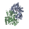
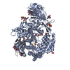
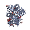
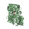
- PDB-4z38: Crystal structure of enoyl reductase domain of MlnA from the macr... -
+ Open data

- Basic information
| Entry | Database: PDB / ID: 4z38 | ||||||
|---|---|---|---|---|---|---|---|
| Title | Crystal structure of enoyl reductase domain of MlnA from the macrolactin biosynthesis cluster from Bacillus amyloliquefaciens | ||||||
 Components Components | MlnA | ||||||
 Keywords Keywords | TRANSFERASE / polyketide / pks / enoyl reductase / trans-AT PKS / AT-less / TIM barrel | ||||||
| Function / homology |  Function and homology information Function and homology information[acyl-carrier-protein] S-malonyltransferase / [acyl-carrier-protein] S-malonyltransferase activity / fatty acid biosynthetic process / nucleotide binding / cytosol Similarity search - Function | ||||||
| Biological species | Bacillus amyloliquefaciens subsp. plantarum | ||||||
| Method |  X-RAY DIFFRACTION / SYNCHROTRON / MOLECULAR REPLACEMENT / Resolution: 2.8 Å X-RAY DIFFRACTION / SYNCHROTRON / MOLECULAR REPLACEMENT / Resolution: 2.8 Å | ||||||
 Authors Authors | Jakob, R.P. / Martin, S.F. / Herbst, D.A. / Maier, T. | ||||||
 Citation Citation | Journal: To Be Published Title: The structural organization of trans-AT polyketide synthases: ketoacyl synthase and trans-acting enoyl reductase Authors: Martin, S.F. / Jakob, R.P. / Herbst, D.A. / Maier, T. | ||||||
| History |
|
- Structure visualization
| Structure viewer | Molecule: MolmilJmol/JSmol |
|---|
- Downloads & links
-Download
| PDBx/mmCIF format | 4z38.cif.gz | 514.4 KB | Display | PDBx/mmCIF format |
|---|---|---|---|---|
| PDB format | pdb4z38.ent.gz | 437.6 KB | Display | PDB format |
| PDBx/mmJSON format | 4z38.json.gz | Tree view | PDBx/mmJSON format | |
| Others |  Other downloads Other downloads |
-Validation report
| Arichive directory | https://data.pdbj.org/pub/pdb/validation_reports/z3/4z38ftp://data.pdbj.org/pub/pdb/validation_reports/z3/4z38 | HTTPS FTP |
|---|
-Related structure data
| Related structure data |  4z37C 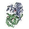 2z6jS S: Starting model for refinement C: citing same article ( |
|---|---|
| Similar structure data |


-Links
 PDBj
PDBj
- Assembly
| Deposited unit | 
| ||||||||
|---|---|---|---|---|---|---|---|---|---|
| 1 |
| ||||||||
| Unit cell |
|
-Components
| #1: Protein | Mass: 53072.551 Da / Num. of mol.: 2 Source method: isolated from a genetically manipulated source Source: (gene. exp.)  Strain: DSM 23117 / BGSC 10A6 / FZB42 / Gene: mlnA, RBAM_014330 / Plasmid: pNIC28a-Bsa4 / Production host: #2: Chemical |   Mass: 456.344 Da / Num. of mol.: 2 / Source method: isolated from a natural source / Formula: C17H21N4O9P Mass: 456.344 Da / Num. of mol.: 2 / Source method: isolated from a natural source / Formula: C17H21N4O9P#3: Water | ChemComp-HOH / |  Mass: 18.015 Da / Num. of mol.: 113 / Source method: isolated from a natural source / Formula: H2O Mass: 18.015 Da / Num. of mol.: 113 / Source method: isolated from a natural source / Formula: H2O |
|---|
-Experimental details
-Experiment
| Experiment | Method: X-RAY DIFFRACTION |
|---|
- Sample preparation
| Crystal | Density Matthews: 2.22 Å3/Da / Density % sol: 44.48 % |
|---|---|
| Crystal grow | Temperature: 293 K / Method: vapor diffusion, sitting drop / Details: 15 % PEG3350, 0.2 M sodium malonate, pH 7 |
-Data collection
| Diffraction | Mean temperature: 100 K |
|---|---|
| Diffraction source | Source: SYNCHROTRON / Site: SLS  / Beamline: X06DA / Wavelength: 1.00003 Å / Beamline: X06DA / Wavelength: 1.00003 Å |
| Detector | Type: DECTRIS PILATUS 2M / Detector: PIXEL / Date: Feb 28, 2013 |
| Radiation | Protocol: SINGLE WAVELENGTH / Monochromatic (M) / Laue (L): M / Scattering type: x-ray |
| Radiation wavelength | Wavelength: 1.00003 Å / Relative weight: 1 |
| Reflection | Resolution: 2.8→108.05 Å / Num. obs: 23587 / % possible obs: 99.6 % / Redundancy: 6.5 % / Biso Wilson estimate: 95.35 Å2 / Rmerge(I) obs: 0.068 / Net I/σ(I): 18.8 |
| Reflection shell | Resolution: 2.8→2.92 Å / Rmerge(I) obs: 0.7 / Mean I/σ(I) obs: 1.7 / % possible all: 99 |
- Processing
| Software |
| ||||||||||||||||||||||||||||||||||||||||||||||||||||||||||||||||||||||||||||||||||||||||||||||||||||||||||||||||||
|---|---|---|---|---|---|---|---|---|---|---|---|---|---|---|---|---|---|---|---|---|---|---|---|---|---|---|---|---|---|---|---|---|---|---|---|---|---|---|---|---|---|---|---|---|---|---|---|---|---|---|---|---|---|---|---|---|---|---|---|---|---|---|---|---|---|---|---|---|---|---|---|---|---|---|---|---|---|---|---|---|---|---|---|---|---|---|---|---|---|---|---|---|---|---|---|---|---|---|---|---|---|---|---|---|---|---|---|---|---|---|---|---|---|---|---|
| Refinement | Method to determine structure: MOLECULAR REPLACEMENT Starting model: 2Z6J Resolution: 2.8→108.05 Å / Cor.coef. Fo:Fc: 0.8982 / Cor.coef. Fo:Fc free: 0.8992 / Cross valid method: THROUGHOUT / σ(F): 0 / SU Rfree Blow DPI: 0.396
| ||||||||||||||||||||||||||||||||||||||||||||||||||||||||||||||||||||||||||||||||||||||||||||||||||||||||||||||||||
| Displacement parameters | Biso mean: 120.43 Å2
| ||||||||||||||||||||||||||||||||||||||||||||||||||||||||||||||||||||||||||||||||||||||||||||||||||||||||||||||||||
| Refine analyze | Luzzati coordinate error obs: 0.849 Å | ||||||||||||||||||||||||||||||||||||||||||||||||||||||||||||||||||||||||||||||||||||||||||||||||||||||||||||||||||
| Refinement step | Cycle: LAST / Resolution: 2.8→108.05 Å
| ||||||||||||||||||||||||||||||||||||||||||||||||||||||||||||||||||||||||||||||||||||||||||||||||||||||||||||||||||
| Refine LS restraints |
| ||||||||||||||||||||||||||||||||||||||||||||||||||||||||||||||||||||||||||||||||||||||||||||||||||||||||||||||||||
| LS refinement shell | Resolution: 2.8→2.92 Å / Total num. of bins used: 12
| ||||||||||||||||||||||||||||||||||||||||||||||||||||||||||||||||||||||||||||||||||||||||||||||||||||||||||||||||||
| Refinement TLS params. | T12: 0.152 Å2 / Method: refined / Refine-ID: X-RAY DIFFRACTION
| ||||||||||||||||||||||||||||||||||||||||||||||||||||||||||||||||||||||||||||||||||||||||||||||||||||||||||||||||||
| Refinement TLS group |
|
