- PDB-4yu8: Crystal structure of Neuroblastoma suppressor of tumorigenicity 1 -
+
Open data
ID or keywords:
Loading...
-
Basic information
Entry
Database: PDB / ID: 4yu8
Title
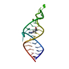
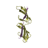
Crystal structure of Neuroblastoma suppressor of tumorigenicity 1
Components
Neuroblastoma suppressor of tumorigenicity 1
Keywords
ANTITUMOR PROTEIN / BMP Binding Protein / NBL1 DAN domain family member 1 BMP-binding / Structural Genomics / Oxford Protein Production Facility / OPPF
Function / homology
Function and homology information
: / negative regulation of monocyte chemotaxis / determination of dorsal identity / morphogen activity / BMP binding / receptor ligand inhibitor activity / negative regulation of BMP signaling pathway / neuron projection morphogenesis / positive regulation of neuron differentiation / protein sequestering activity ...: / negative regulation of monocyte chemotaxis / determination of dorsal identity / morphogen activity / BMP binding / receptor ligand inhibitor activity / negative regulation of BMP signaling pathway / neuron projection morphogenesis / positive regulation of neuron differentiation / protein sequestering activity / nervous system development / receptor ligand activity / : / identical protein binding Similarity search - Function
Neuroblastoma suppressor of tumourigenicity 1 / DAN / DAN domain / Cystine knot, C-terminal / C-terminal cystine knot-like domain (CTCK) / Cystine-knot cytokine Similarity search - Domain/homology
Neuroblastomasuppressoroftumorigenicity1 / DAN domain family member 1 / Protein N03 / Zinc finger protein DAN
Mass: 15110.457 Da / Num. of mol.: 1 Source method: isolated from a genetically manipulated source Source: (gene. exp.) Homo sapiens (human) / Gene: NBL1, DAN, DAND1 / Cell line (production host): HEK293T / Production host: HOMO SAPIENS (human) / References: UniProt: P41271
Resolution: 1.8→42.62 Å / Cor.coef. Fo:Fc: 0.957 / Cor.coef. Fo:Fc free: 0.943 / SU B: 8.111 / SU ML: 0.103 / Cross valid method: THROUGHOUT / ESU R: 0.119 / ESU R Free: 0.123 / Stereochemistry target values: MAXIMUM LIKELIHOOD / Details: HYDROGENS HAVE BEEN ADDED IN THE RIDING POSITIONS
Rfactor
Num. reflection
% reflection
Selection details
Rfree
0.25778
479
4.8 %
RANDOM
Rwork
0.21411
-
-
-
obs
0.21605
9496
99.22 %
-
Solvent computation
Ion probe radii: 0.8 Å / Shrinkage radii: 0.8 Å / VDW probe radii: 1.2 Å / Solvent model: MASK
 Movie
Movie Controller

 Yorodumi
 Open data
 Basic information
 Components
 Keywords
 Function and homology information
 Homo sapiens (human)
 X-RAY DIFFRACTION /
  Authors
 Citation
 Structure visualization
 Downloads & links
 Other downloads
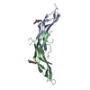


 PDBj
PDBj

 Assembly

 Mass: 92.094 Da / Num. of mol.: 1 / Source method: obtained synthetically / Formula: C3H8O3
 Type: D-saccharide, beta linking / Mass: 221.208 Da / Num. of mol.: 1
 Mass: 18.015 Da / Num. of mol.: 50 / Source method: isolated from a natural source / Formula: H2O
 Sample preparation
 / Beamline: BM14 / Wavelength: 1.278 Å
 Processing