[English] 日本語
Yorodumi
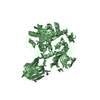
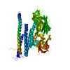
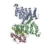
- PDB-4yu5: Crystal structure of selenomethionine variant of Bacillus anthrac... -

+
Open data


ID or keywords:

Loading...

-
Basic information

Entry
Database: PDB / ID: 4yu5
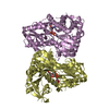
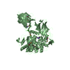
TitleCrystal structure of selenomethionine variant of Bacillus anthracis immune inhibitor A2 peptidase zymogen
ComponentsImmune inhibitor A, metalloprotease
KeywordsHYDROLASE / metallopeptidase / metzincin
Function / homology
Function and homology information


Hydrolases; Acting on peptide bonds (peptidases); Metalloendopeptidases / metallopeptidase activity / proteolysis / metal ion binding
Similarity search - Function
Peptidase M6, InhA / Peptidase M6-like, domain / Immune inhibitor A peptidase M6, catalytic domain / Prokaryotic membrane lipoprotein lipid attachment site profile.
Similarity search - Domain/homology
: / 3-(1-methylpiperidinium-1-yl)propane-1-sulfonate / Immune inhibitor A, metalloprotease
Similarity search - Component
Biological speciesBacillus cereus var. anthracis
MethodX-RAY DIFFRACTION / SYNCHROTRON / MAD / Resolution: 2.9 Å
AuthorsArolas, J.L. / Goulas, T. / Gomis-Ruth, F.X.
CitationJournal: Structure / Year: 2016
Title: Structural Basis for Latency and Function of Immune Inhibitor A Metallopeptidase, a Modulator of the Bacillus anthracis Secretome.
Authors: Arolas, J.L. / Goulas, T. / Pomerantsev, A.P. / Leppla, S.H. / Gomis-Ruth, F.X.
History
DepositionMar 18, 2015Deposition site: RCSB / Processing site: PDBE
Revision 1.0Oct 28, 2015Provider: repository / Type: Initial release
Revision 1.1Jan 20, 2016Group: Database references

-
Structure visualization

Structure viewerMolecule:
MolmilJmol/JSmol

Downloads & links

-
Assembly

Deposited unit
A: Immune inhibitor A, metalloprotease
B: Immune inhibitor A, metalloprotease
hetero molecules


Theoretical massNumber of molelcules
Total (without water)169,73521
Polymers168,2492
Non-polymers1,48619
Water1,874104
1
A: Immune inhibitor A, metalloprotease
hetero molecules


Theoretical massNumber of molelcules
Total (without water)85,34514
Polymers84,1251
Non-polymers1,22113
Water181
TypeNameSymmetry operationNumber
identity operation1_555x,y,z1
2
B: Immune inhibitor A, metalloprotease
hetero molecules


Theoretical massNumber of molelcules
Total (without water)84,3897
Polymers84,1251
Non-polymers2656
Water181
TypeNameSymmetry operationNumber
identity operation1_555x,y,z1
Unit cell
Length a, b, c (Å)97.610, 102.410, 242.880
Angle α, β, γ (deg.)90.00, 90.00, 90.00
Int Tables number19
Space group name H-MP212121

-
Components

-
Protein , 1 types, 2 molecules AB

#1: Protein Immune inhibitor A, metalloprotease


Mass: 84124.500 Da / Num. of mol.: 2 / Fragment: residues 50-799 / Mutation: E331A
Source method: isolated from a genetically manipulated source
Details: The conflicts are due to the uniprot entry being for a different strain
Source: (gene. exp.) Bacillus cereus var. anthracis (strain CI) (bacteria)
Strain: CI / Gene: inhA2, BACI_c06810 / Plasmid: pHT43 / Production host: Bacillus subtilis (bacteria) / Strain (production host): WB800N
References: UniProt: D8H130, Hydrolases; Acting on peptide bonds (peptidases); Metalloendopeptidases

-
Non-polymers , 6 types, 123 molecules

#2: Chemical ChemComp-ZN / ZINC ION


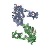
Mass: 65.409 Da / Num. of mol.: 2 / Source method: obtained synthetically / Formula: Zn
#3: Chemical
ChemComp-CA / CALCIUM ION


Mass: 40.078 Da / Num. of mol.: 8 / Source method: obtained synthetically / Formula: Ca
#4: Chemical ChemComp-KH2 / 3-(1-methylpiperidinium-1-yl)propane-1-sulfonate


Mass: 221.317 Da / Num. of mol.: 2 / Source method: obtained synthetically / Formula: C9H19NO3S
#5: Chemical
ChemComp-GOL / GLYCEROL / GLYCERIN / PROPANE-1,2,3-TRIOL


Mass: 92.094 Da / Num. of mol.: 6 / Source method: obtained synthetically / Formula: C3H8O3
#6: Chemical ChemComp-K / POTASSIUM ION


Mass: 39.098 Da / Num. of mol.: 1 / Source method: obtained synthetically / Formula: K
#7: Water ChemComp-HOH / water


Mass: 18.015 Da / Num. of mol.: 104 / Source method: isolated from a natural source / Formula: H2O

-
Experimental details

-
Experiment

ExperimentMethod: X-RAY DIFFRACTION

-
Sample preparation

CrystalDensity Matthews: 3.61 Å3/Da / Density % sol: 65.91 %
Crystal growTemperature: 277 K / Method: vapor diffusion, hanging drop / pH: 7.5
Details: 0.1 M BIS-TRIS propane, 0.2 M potassium sodium tartrate, 20% (w/v) PEG 3350

-
Data collection

DiffractionMean temperature: 100 K
Diffraction sourceSource: SYNCHROTRON / Site: ALBA / Beamline: XALOC / Wavelength: 0.97926, 0.93923
DetectorType: DECTRIS PILATUS 6M / Detector: PIXEL / Date: Mar 14, 2014
RadiationMonochromator: Si(111) / Protocol: MAD / Monochromatic (M) / Laue (L): M / Scattering type: x-ray
Radiation wavelength
IDWavelength (Å)Relative weight
10.979261
20.939231
ReflectionResolution: 2.9→48.8 Å / Num. obs: 54813 / % possible obs: 99.8 % / Redundancy: 8.4 % / Biso Wilson estimate: 74.38 Å2 / Rmerge(I) obs: 0.114 / Net I/σ(I): 15.2
Reflection shellResolution: 2.9→3.07 Å / Redundancy: 8.5 % / Rmerge(I) obs: 0.672 / Mean I/σ(I) obs: 4.2 / % possible all: 99.8

-
Processing

Software
NameVersionClassification
XDSdata reduction
XSCALEdata scaling
Cootmodel building
SHELXphasing
BUSTER2.11.5refinement
RefinementMethod to determine structure: MAD / Resolution: 2.9→42.46 Å / Cor.coef. Fo:Fc: 0.9105 / Cor.coef. Fo:Fc free: 0.9075 / SU R Cruickshank DPI: 0.529 / Cross valid method: THROUGHOUT / σ(F): 0 / SU R Blow DPI: 0.5 / SU Rfree Blow DPI: 0.261 / SU Rfree Cruickshank DPI: 0.268
RfactorNum. reflection% reflectionSelection details
Rfree0.2071 706 1.29 %RANDOM
Rwork0.1824 ---
obs0.1827 54706 99.86 %-
Displacement parametersBiso mean: 71.82 Å2
Baniso -1Baniso -2Baniso -3
1-6.4017 Å20 Å20 Å2
2---25.8688 Å20 Å2
3---19.467 Å2
Refine analyzeLuzzati coordinate error obs: 0.438 Å
Refinement stepCycle: 1 / Resolution: 2.9→42.46 Å
ProteinNucleic acidLigandSolventTotal
Num. atoms10763 0 75 104 10942
Refine LS restraints
Refine-IDTypeDev idealNumberRestraint functionWeight
X-RAY DIFFRACTIONt_bond_d0.0111078HARMONIC2
X-RAY DIFFRACTIONt_angle_deg1.1414973HARMONIC2
X-RAY DIFFRACTIONt_dihedral_angle_d5007SINUSOIDAL2
X-RAY DIFFRACTIONt_incorr_chiral_ct
X-RAY DIFFRACTIONt_pseud_angle
X-RAY DIFFRACTIONt_trig_c_planes311HARMONIC2
X-RAY DIFFRACTIONt_gen_planes1599HARMONIC5
X-RAY DIFFRACTIONt_it11078HARMONIC20
X-RAY DIFFRACTIONt_nbd2SEMIHARMONIC5
X-RAY DIFFRACTIONt_omega_torsion3.28
X-RAY DIFFRACTIONt_other_torsion3.21
X-RAY DIFFRACTIONt_improper_torsion
X-RAY DIFFRACTIONt_chiral_improper_torsion1362SEMIHARMONIC5
X-RAY DIFFRACTIONt_sum_occupancies
X-RAY DIFFRACTIONt_utility_distance
X-RAY DIFFRACTIONt_utility_angle
X-RAY DIFFRACTIONt_utility_torsion
X-RAY DIFFRACTIONt_ideal_dist_contact12043SEMIHARMONIC4
LS refinement shellResolution: 2.9→2.98 Å / Total num. of bins used: 20
RfactorNum. reflection% reflection
Rfree0.3637 50 1.26 %
Rwork0.2415 3917 -
all0.2429 3967 -
obs--99.86 %
Refinement TLS params.

Method: refined / Refine-ID: X-RAY DIFFRACTION

IDL112)L122)L132)L222)L232)L332)S11 (Å °)S12 (Å °)S13 (Å °)S21 (Å °)S22 (Å °)S23 (Å °)S31 (Å °)S32 (Å °)S33 (Å °)T112)T122)T132)T222)T232)T332)Origin x (Å)Origin y (Å)Origin z (Å)
11.2482-0.12640.18541.50040.99762.62750.13580.2892-0.1899-0.11560.0933-0.0757-0.12650.0254-0.2291-0.1532-0.01880.0005-0.0884-0.05-0.122483.879442.1481249.355
23.4851.6277-2.00692.7758-2.11418.1155-0.0064-0.03-0.27090.17230.15010.3590.6357-0.5908-0.14370.19-0.12930.1385-0.11840.01690.035196.241973.9219221.201
34.85960.82322.249901.62694.6003-0.05450.24730.21840.02760.3758-0.2405-0.06270.2392-0.3213-0.13910.0714-0.08020.1245-0.26710.3759103.21932.4421245.098
41.39050.12860.33241.6845-0.07273.0222-0.1433-0.4216-0.20030.64490.2387-0.23460.3695-0.0775-0.09540.34580.2033-0.0544-0.08810.0107-0.09986.255324.1315181.177
54.39510.6725-3.19212.3609-0.17246.0220.06490.16230.386-0.29340.1797-0.3163-0.37080.3622-0.24460.1723-0.11790.0755-0.06070.0453-0.021647.266914.1304199.106
60-1.532-0.42865.9498-1.6347.0394-0.015-0.0428-0.2158-0.0262-0.0272-0.1290.11270.01340.04220.58240.2749-0.117-0.0815-0.00330.335697.22153.3767181.354
Refinement TLS group
IDRefine-IDRefine TLS-IDSelection details
1X-RAY DIFFRACTION1{A|144 - 482 A|642 - 799 A|901 - 903 A|995 - 999}
2X-RAY DIFFRACTION2{A|483 - 641}
3X-RAY DIFFRACTION3{A|50 - 94}
4X-RAY DIFFRACTION4{B|144 - 482 B|642 - 799 B|901 - 902 B|995 - 999}
5X-RAY DIFFRACTION5{B|483 - 641}
6X-RAY DIFFRACTION6{B|50 - 89}

+
About Yorodumi

-
News

-
Feb 9, 2022. New format data for meta-information of EMDB entries

New format data for meta-information of EMDB entries

  • Version 3 of the EMDB header file is now the official format.
  • The previous official version 1.9 will be removed from the archive.

Related info.:EMDB header

External links:wwPDB to switch to version 3 of the EMDB data model

-
Aug 12, 2020. Covid-19 info

Covid-19 info

URL: https://pdbjlvh1.pdbj.org/emnavi/covid19.php

New page: Covid-19 featured information page in EM Navigator.

Related info.:Covid-19 info / Mar 5, 2020. Novel coronavirus structure data

+
Mar 5, 2020. Novel coronavirus structure data

Novel coronavirus structure data

Related info.:Yorodumi Speices / Aug 12, 2020. Covid-19 info

External links:COVID-19 featured content - PDBj / Molecule of the Month (242):Coronavirus Proteases

+
Jan 31, 2019. EMDB accession codes are about to change! (news from PDBe EMDB page)

EMDB accession codes are about to change! (news from PDBe EMDB page)

  • The allocation of 4 digits for EMDB accession codes will soon come to an end. Whilst these codes will remain in use, new EMDB accession codes will include an additional digit and will expand incrementally as the available range of codes is exhausted. The current 4-digit format prefixed with “EMD-” (i.e. EMD-XXXX) will advance to a 5-digit format (i.e. EMD-XXXXX), and so on. It is currently estimated that the 4-digit codes will be depleted around Spring 2019, at which point the 5-digit format will come into force.
  • The EM Navigator/Yorodumi systems omit the EMD- prefix.

Related info.:Q: What is EMD? / ID/Accession-code notation in Yorodumi/EM Navigator

External links:EMDB Accession Codes are Changing Soon! / Contact to PDBj

+
Jul 12, 2017. Major update of PDB

Major update of PDB

  • wwPDB released updated PDB data conforming to the new PDBx/mmCIF dictionary.
  • This is a major update changing the version number from 4 to 5, and with Remediation, in which all the entries are updated.
  • In this update, many items about electron microscopy experimental information are reorganized (e.g. em_software).
  • Now, EM Navigator and Yorodumi are based on the updated data.

External links:wwPDB Remediation / Enriched Model Files Conforming to OneDep Data Standards Now Available in the PDB FTP Archive

-
Yorodumi

Thousand views of thousand structures

  • Yorodumi is a browser for structure data from EMDB, PDB, SASBDB, etc.
  • This page is also the successor to EM Navigator detail page, and also detail information page/front-end page for Omokage search.
  • The word "yorodu" (or yorozu) is an old Japanese word meaning "ten thousand". "mi" (miru) is to see.

Related info.:EMDB / PDB / SASBDB / Comparison of 3 databanks / Yorodumi Search / Aug 31, 2016. New EM Navigator & Yorodumi / Yorodumi Papers / Jmol/JSmol / Function and homology information / Changes in new EM Navigator and Yorodumi

Read more