 Movie
Movie Controller

[English] 日本語
 Yorodumi
- PDB-4yq1: Crystal structure of TrmD, a M1G37 tRNA Methyltransferase with SA... -
+ Open data

- Basic information
| Entry | Database: PDB / ID: 4yq1 | ||||||
|---|---|---|---|---|---|---|---|
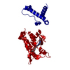
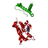
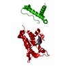
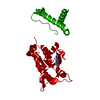
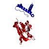
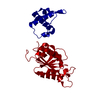
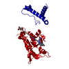
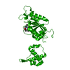
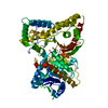
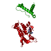
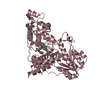
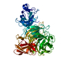
| Title | Crystal structure of TrmD, a M1G37 tRNA Methyltransferase with SAM-competitive compounds | ||||||
 Components Components | tRNA (guanine-N(1)-)-methyltransferase | ||||||
 Keywords Keywords | transferase/transferase inhibitor / TrmD / SAM-binding / knot / transferase-transferase inhibitor complex | ||||||
| Function / homology |  Function and homology information Function and homology informationtRNA (guanine37-N1)-methyltransferase / tRNA (guanine(37)-N1)-methyltransferase activity / tRNA N1-guanine methylation / cytosol Similarity search - Function | ||||||
| Biological species |  Haemophilus influenzae (bacteria) Haemophilus influenzae (bacteria) | ||||||
| Method |  X-RAY DIFFRACTION / SYNCHROTRON / MOLECULAR REPLACEMENT / Resolution: 2 Å X-RAY DIFFRACTION / SYNCHROTRON / MOLECULAR REPLACEMENT / Resolution: 2 Å | ||||||
 Authors Authors | Elkins, P.A. / Bonnette, W.G. / Madauss, K.P. / Stuckey, J.A. | ||||||
 Citation Citation | Journal: To be published Title: To Be Published Authors: Elkins, P.A. / Bonnette, W.G. / Madauss, K.P. / Stuckey, J.A. | ||||||
| History |
|
- Structure visualization
| Structure viewer | Molecule: MolmilJmol/JSmol |
|---|
- Downloads & links
-Download
| PDBx/mmCIF format | 4yq1.cif.gz | 116.1 KB | Display | PDBx/mmCIF format |
|---|---|---|---|---|
| PDB format | pdb4yq1.ent.gz | 88 KB | Display | PDB format |
| PDBx/mmJSON format | 4yq1.json.gz | Tree view | PDBx/mmJSON format | |
| Others |  Other downloads Other downloads |
-Validation report
| Arichive directory | https://data.pdbj.org/pub/pdb/validation_reports/yq/4yq1ftp://data.pdbj.org/pub/pdb/validation_reports/yq/4yq1 | HTTPS FTP |
|---|
-Related structure data
| Related structure data |  1p9pS S: Starting model for refinement |
|---|---|
| Similar structure data |


-Links
 PDBj
PDBj- Assembly
| Deposited unit | 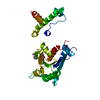
| |||||||||
|---|---|---|---|---|---|---|---|---|---|---|
| 1 | 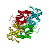
| |||||||||
| 2 | 
| |||||||||
| Unit cell |
| |||||||||
| Components on special symmetry positions |
|
-Components
| #1: Protein | Mass: 28384.576 Da / Num. of mol.: 1 Source method: isolated from a genetically manipulated source Source: (gene. exp.) Haemophilus influenzae (strain ATCC 51907 / DSM 11121 / KW20 / Rd) (bacteria)Strain: ATCC 51907 / DSM 11121 / KW20 / Rd / Gene: trmD, HI_0202 / Plasmid: pET28a / Production host: References: UniProt: P43912, tRNA (guanine37-N1)-methyltransferase |
|---|---|
| #2: Chemical | ChemComp-4FN /   Mass: 433.543 Da / Num. of mol.: 1 / Source method: obtained synthetically / Formula: C26H31N3O3 Mass: 433.543 Da / Num. of mol.: 1 / Source method: obtained synthetically / Formula: C26H31N3O3 |
| #3: Water | ChemComp-HOH /  Mass: 18.015 Da / Num. of mol.: 84 / Source method: isolated from a natural source / Formula: H2O Mass: 18.015 Da / Num. of mol.: 84 / Source method: isolated from a natural source / Formula: H2O |
-Experimental details
-Experiment
| Experiment | Method: X-RAY DIFFRACTION / Number of used crystals: 1 |
|---|
- Sample preparation
| Crystal | Density Matthews: 2.85 Å3/Da / Density % sol: 56.77 % |
|---|---|
| Crystal grow | Temperature: 295 K / Method: vapor diffusion, sitting drop / pH: 7.5 Details: Protein solution:( 12/mg/mL in 100mM HEPES pH 7.5, 150mM NaCl, 10mM MgCl2 2mM DTT) Well solution: (20% PEG3,350 and 0.2M potassium citrate tribasic monohydrate). 4uL of S-adenosyl methionine ...Details: Protein solution:( 12/mg/mL in 100mM HEPES pH 7.5, 150mM NaCl, 10mM MgCl2 2mM DTT) Well solution: (20% PEG3,350 and 0.2M potassium citrate tribasic monohydrate). 4uL of S-adenosyl methionine in water were added to 100uL of protein and allowed to incubate on ice for 1 hour before protein was mixed with well at 1:1 ratio.Seeding used to improve crystals. Compound stock solutions (either 100mM or 1M stocks) were added up to a final drop concentration of 4.8% DMSO. Crystals were soaked for 4-6 hours. 20% glycerol in well solution was used as cryoprotectant for a quick dip of crystal in liquid N2. |
-Data collection
| Diffraction | Mean temperature: 100 K | ||||||||||||||||||||||||||||||||||||||||||||||||||||||||||||||||||
|---|---|---|---|---|---|---|---|---|---|---|---|---|---|---|---|---|---|---|---|---|---|---|---|---|---|---|---|---|---|---|---|---|---|---|---|---|---|---|---|---|---|---|---|---|---|---|---|---|---|---|---|---|---|---|---|---|---|---|---|---|---|---|---|---|---|---|---|
| Diffraction source | Source: SYNCHROTRON / Site: APS  / Beamline: 21-ID-F / Wavelength: 0.97872 Å / Beamline: 21-ID-F / Wavelength: 0.97872 Å | ||||||||||||||||||||||||||||||||||||||||||||||||||||||||||||||||||
| Detector | Type: MARMOSAIC 300 mm CCD / Detector: CCD / Date: Nov 4, 2009 | ||||||||||||||||||||||||||||||||||||||||||||||||||||||||||||||||||
| Radiation | Monochromator: unknown / Protocol: SINGLE WAVELENGTH / Monochromatic (M) / Laue (L): M / Scattering type: x-ray | ||||||||||||||||||||||||||||||||||||||||||||||||||||||||||||||||||
| Radiation wavelength | Wavelength: 0.97872 Å / Relative weight: 1 | ||||||||||||||||||||||||||||||||||||||||||||||||||||||||||||||||||
| Reflection | Resolution: 2→50 Å / Num. obs: 22253 / % possible obs: 99.9 % / Redundancy: 7.4 % / Biso Wilson estimate: 28.72 Å2 / Rmerge(I) obs: 0.077 / Χ2: 0.927 / Net I/av σ(I): 6.862 / Net I/σ(I): 12.2 / Num. measured all: 163780 | ||||||||||||||||||||||||||||||||||||||||||||||||||||||||||||||||||
| Reflection shell | Diffraction-ID: 1 / Rejects: _
|
- Processing
| Software |
| |||||||||||||||||||||||||||||||||||||||||||||||||||||||||||||||||||||||||||||||||||||||||||||||||||||||||
|---|---|---|---|---|---|---|---|---|---|---|---|---|---|---|---|---|---|---|---|---|---|---|---|---|---|---|---|---|---|---|---|---|---|---|---|---|---|---|---|---|---|---|---|---|---|---|---|---|---|---|---|---|---|---|---|---|---|---|---|---|---|---|---|---|---|---|---|---|---|---|---|---|---|---|---|---|---|---|---|---|---|---|---|---|---|---|---|---|---|---|---|---|---|---|---|---|---|---|---|---|---|---|---|---|---|---|
| Refinement | Method to determine structure: MOLECULAR REPLACEMENT Starting model: 1P9P Resolution: 2→31.396 Å / FOM work R set: 0.8522 / SU ML: 0.17 / Cross valid method: THROUGHOUT / σ(F): 1.34 / Phase error: 21.7 / Stereochemistry target values: ML
| |||||||||||||||||||||||||||||||||||||||||||||||||||||||||||||||||||||||||||||||||||||||||||||||||||||||||
| Solvent computation | Shrinkage radii: 0.9 Å / VDW probe radii: 1.11 Å / Solvent model: FLAT BULK SOLVENT MODEL | |||||||||||||||||||||||||||||||||||||||||||||||||||||||||||||||||||||||||||||||||||||||||||||||||||||||||
| Displacement parameters | Biso max: 115.96 Å2 / Biso mean: 35.81 Å2 / Biso min: 14.93 Å2 | |||||||||||||||||||||||||||||||||||||||||||||||||||||||||||||||||||||||||||||||||||||||||||||||||||||||||
| Refinement step | Cycle: final / Resolution: 2→31.396 Å
| |||||||||||||||||||||||||||||||||||||||||||||||||||||||||||||||||||||||||||||||||||||||||||||||||||||||||
| Refine LS restraints |
| |||||||||||||||||||||||||||||||||||||||||||||||||||||||||||||||||||||||||||||||||||||||||||||||||||||||||
| LS refinement shell | Refine-ID: X-RAY DIFFRACTION / Total num. of bins used: 14
| |||||||||||||||||||||||||||||||||||||||||||||||||||||||||||||||||||||||||||||||||||||||||||||||||||||||||
| Refinement TLS params. | Method: refined / Refine-ID: X-RAY DIFFRACTION
| |||||||||||||||||||||||||||||||||||||||||||||||||||||||||||||||||||||||||||||||||||||||||||||||||||||||||
| Refinement TLS group |
|
