 Movie
Movie Controller

[English] 日本語
 Yorodumi
- PDB-3axz: Crystal structure of Haemophilus influenzae TrmD in complex with ... -
+ Open data

- Basic information
| Entry | Database: PDB / ID: 3axz | ||||||
|---|---|---|---|---|---|---|---|
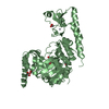
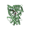
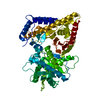
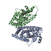
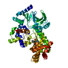
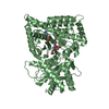
| Title | Crystal structure of Haemophilus influenzae TrmD in complex with adenosine | ||||||
 Components Components | tRNA (guanine-N(1)-)-methyltransferase | ||||||
 Keywords Keywords | TRANSFERASE / trefoil knot / methyltransferase / AdoMet binding | ||||||
| Function / homology |  Function and homology information Function and homology informationtRNA (guanine37-N1)-methyltransferase / tRNA (guanine(37)-N1)-methyltransferase activity / tRNA N1-guanine methylation / cytosol Similarity search - Function | ||||||
| Biological species |  Haemophilus influenzae (bacteria) Haemophilus influenzae (bacteria) | ||||||
| Method |  X-RAY DIFFRACTION / SYNCHROTRON / MOLECULAR REPLACEMENT / Resolution: 2.25 Å X-RAY DIFFRACTION / SYNCHROTRON / MOLECULAR REPLACEMENT / Resolution: 2.25 Å | ||||||
 Authors Authors | Yoshida, K. / Goto-Ito, S. / Ito, T. / Hou, Y.M. / Yokoyama, S. | ||||||
 Citation Citation | Journal: Rna / Year: 2011 Title: Differentiating analogous tRNA methyltransferases by fragments of the methyl donor. Authors: Lahoud, G. / Goto-Ito, S. / Yoshida, K. / Ito, T. / Yokoyama, S. / Hou, Y.M. | ||||||
| History |
|
- Structure visualization
| Structure viewer | Molecule: MolmilJmol/JSmol |
|---|
- Downloads & links
-Download
| PDBx/mmCIF format | 3axz.cif.gz | 64.6 KB | Display | PDBx/mmCIF format |
|---|---|---|---|---|
| PDB format | pdb3axz.ent.gz | 46.5 KB | Display | PDB format |
| PDBx/mmJSON format | 3axz.json.gz | Tree view | PDBx/mmJSON format | |
| Others |  Other downloads Other downloads |
-Validation report
| Arichive directory | https://data.pdbj.org/pub/pdb/validation_reports/ax/3axzftp://data.pdbj.org/pub/pdb/validation_reports/ax/3axz | HTTPS FTP |
|---|
-Related structure data
| Related structure data |  3ay0C  1uamS C: citing same article ( S: Starting model for refinement |
|---|---|
| Similar structure data |

-Links
 PDBj
PDBj
- Assembly
| Deposited unit | 
| ||||||||
|---|---|---|---|---|---|---|---|---|---|
| 1 | 
| ||||||||
| Unit cell |
|
-Components
| #1: Protein | Mass: 29750.016 Da / Num. of mol.: 1 Source method: isolated from a genetically manipulated source Source: (gene. exp.) Haemophilus influenzae (bacteria) / Strain: KW20 / Gene: HI_0202, trmD / Plasmid: pET-28b / Production host: |
|---|---|
| #2: Chemical | ChemComp-ADN /   Mass: 267.241 Da / Num. of mol.: 1 / Source method: obtained synthetically / Formula: C10H13N5O4 Mass: 267.241 Da / Num. of mol.: 1 / Source method: obtained synthetically / Formula: C10H13N5O4 |
| #3: Water | ChemComp-HOH /  Mass: 18.015 Da / Num. of mol.: 121 / Source method: isolated from a natural source / Formula: H2O Mass: 18.015 Da / Num. of mol.: 121 / Source method: isolated from a natural source / Formula: H2O |
-Experimental details
-Experiment
| Experiment | Method: X-RAY DIFFRACTION / Number of used crystals: 1 |
|---|
- Sample preparation
| Crystal | Density Matthews: 2.59 Å3/Da / Density % sol: 52.6 % |
|---|---|
| Crystal grow | Temperature: 298 K / Method: vapor diffusion, sitting drop / pH: 6.5 Details: 0.2M calcium acetate, 0.1M sodium cacodylate, 18% PEG 8000, pH 6.5, VAPOR DIFFUSION, SITTING DROP, temperature 298.0K |
-Data collection
| Diffraction | Mean temperature: 95 K |
|---|---|
| Diffraction source | Source: SYNCHROTRON / Site: Photon Factory  / Beamline: AR-NE3A / Wavelength: 1 Å / Beamline: AR-NE3A / Wavelength: 1 Å |
| Detector | Type: ADSC QUANTUM 270 / Detector: CCD / Date: Jun 16, 2010 |
| Radiation | Monochromator: Numerical link type Si(111) double crystal monochromator Protocol: SINGLE WAVELENGTH / Monochromatic (M) / Laue (L): M / Scattering type: x-ray |
| Radiation wavelength | Wavelength: 1 Å / Relative weight: 1 |
| Reflection | Resolution: 2.25→50 Å / Num. obs: 14775 / % possible obs: 98.7 % / Observed criterion σ(I): -3 / Redundancy: 5.8 % / Biso Wilson estimate: 25 Å2 / Rsym value: 0.152 / Net I/σ(I): 11.2 |
| Reflection shell | Resolution: 2.25→2.29 Å / Redundancy: 4.2 % / Mean I/σ(I) obs: 2.2 / Num. unique all: 668 / Rsym value: 0.427 / % possible all: 91.3 |
- Processing
| Software |
| ||||||||||||||||||||
|---|---|---|---|---|---|---|---|---|---|---|---|---|---|---|---|---|---|---|---|---|---|
| Refinement | Method to determine structure: MOLECULAR REPLACEMENT Starting model: PDB ENTRY 1UAM Resolution: 2.25→50 Å / Cross valid method: THROUGHOUT / σ(F): 0
| ||||||||||||||||||||
| Refinement step | Cycle: LAST / Resolution: 2.25→50 Å
| ||||||||||||||||||||
| Refine LS restraints |
|
