 Movie
Movie Controller

[English] 日本語
 Yorodumi
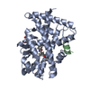
- PDB-4ypq: Crystal structure of the ROR(gamma)t ligand binding domain in com... -
+ Open data

- Basic information
| Entry | Database: PDB / ID: 4ypq | ||||||
|---|---|---|---|---|---|---|---|
| Title | Crystal structure of the ROR(gamma)t ligand binding domain in complex with 4-(1-(2-chloro-6-(trifluoromethyl)benzoyl)-1H-indazol-3-yl)benzoic acid | ||||||
 Components Components | Nuclear receptor ROR-gamma | ||||||
 Keywords Keywords | TRANSCRIPTION / nuclear receptor ligand binding domain | ||||||
| Function / homology |  Function and homology information Function and homology informationtolerance induction in gut-associated lymphoid tissue / regulation of steroid metabolic process / cellular response to sterol / ligand-modulated transcription factor activity / Peyer's patch development / positive regulation of circadian rhythm / regulatory T cell differentiation / T-helper cell differentiation / oxysterol binding / RUNX3 Regulates Immune Response and Cell Migration ...tolerance induction in gut-associated lymphoid tissue / regulation of steroid metabolic process / cellular response to sterol / ligand-modulated transcription factor activity / Peyer's patch development / positive regulation of circadian rhythm / regulatory T cell differentiation / T-helper cell differentiation / oxysterol binding / RUNX3 Regulates Immune Response and Cell Migration / negative regulation of thymocyte apoptotic process / regulation of fat cell differentiation / Phosphorylated BMAL1:CLOCK (ARNTL:CLOCK) activates expression of core clock genes / T-helper 17 cell differentiation / adipose tissue development / regulation of glucose metabolic process / lymph node development / xenobiotic metabolic process / RORA,B,C and NR1D1 (REV-ERBA) regulate gene expression / Expression of BMAL (ARNTL), CLOCK, and NPAS2 / circadian regulation of gene expression / Nuclear Receptor transcription pathway / DNA-binding transcription repressor activity, RNA polymerase II-specific / nuclear receptor activity / sequence-specific double-stranded DNA binding / Interleukin-4 and Interleukin-13 signaling / DNA-binding transcription factor activity, RNA polymerase II-specific / nuclear body / RNA polymerase II cis-regulatory region sequence-specific DNA binding / DNA-binding transcription factor activity / regulation of transcription by RNA polymerase II / positive regulation of DNA-templated transcription / chromatin / negative regulation of transcription by RNA polymerase II / zinc ion binding / nucleoplasm / nucleus Similarity search - Function | ||||||
| Biological species |  Homo sapiens (human) Homo sapiens (human) | ||||||
| Method |  X-RAY DIFFRACTION / SYNCHROTRON / MOLECULAR REPLACEMENT / Resolution: 2.32 Å X-RAY DIFFRACTION / SYNCHROTRON / MOLECULAR REPLACEMENT / Resolution: 2.32 Å | ||||||
 Authors Authors | Leysen, S. / Scheepstra, M. / van Almen, G.C. / Ottmann, C. / Brunsveld, L. | ||||||
 Citation Citation | Journal: Nat Commun / Year: 2015 Title: Identification of an allosteric binding site for ROR gamma t inhibition. Authors: Scheepstra, M. / Leysen, S. / van Almen, G.C. / Miller, J.R. / Piesvaux, J. / Kutilek, V. / van Eenennaam, H. / Zhang, H. / Barr, K. / Nagpal, S. / Soisson, S.M. / Kornienko, M. / Wiley, K. ...Authors: Scheepstra, M. / Leysen, S. / van Almen, G.C. / Miller, J.R. / Piesvaux, J. / Kutilek, V. / van Eenennaam, H. / Zhang, H. / Barr, K. / Nagpal, S. / Soisson, S.M. / Kornienko, M. / Wiley, K. / Elsen, N. / Sharma, S. / Correll, C.C. / Trotter, B.W. / van der Stelt, M. / Oubrie, A. / Ottmann, C. / Parthasarathy, G. / Brunsveld, L. | ||||||
| History |
|
- Structure visualization
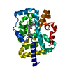
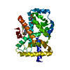
| Structure viewer | Molecule: MolmilJmol/JSmol |
|---|
- Downloads & links
-Download
| PDBx/mmCIF format | 4ypq.cif.gz | 67.8 KB | Display | PDBx/mmCIF format |
|---|---|---|---|---|
| PDB format | pdb4ypq.ent.gz | 49 KB | Display | PDB format |
| PDBx/mmJSON format | 4ypq.json.gz | Tree view | PDBx/mmJSON format | |
| Others |  Other downloads Other downloads |
-Validation report
| Arichive directory | https://data.pdbj.org/pub/pdb/validation_reports/yp/4ypqftp://data.pdbj.org/pub/pdb/validation_reports/yp/4ypq | HTTPS FTP |
|---|
-Related structure data
| Related structure data | 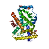 5c4oC  5c4sC 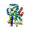 5c4tC 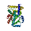 5c4uC  4nieS S: Starting model for refinement C: citing same article ( |
|---|---|
| Similar structure data |


-Links
 PDBj
PDBj- Assembly
| Deposited unit | 
| ||||||||||||||||||||||||
|---|---|---|---|---|---|---|---|---|---|---|---|---|---|---|---|---|---|---|---|---|---|---|---|---|---|
| 1 |
| ||||||||||||||||||||||||
| Unit cell |
| ||||||||||||||||||||||||
| Components on special symmetry positions |
|
-Components
| #1: Protein | Mass: 28307.824 Da / Num. of mol.: 1 / Fragment: UNP residues 265-507 Source method: isolated from a genetically manipulated source Source: (gene. exp.) Homo sapiens (human) / Gene: RORC, NR1F3, RORG, RZRG / Production host:  |
|---|---|
| #2: Chemical | ChemComp-4F1 /   Mass: 444.790 Da / Num. of mol.: 1 / Source method: obtained synthetically / Formula: C22H12ClF3N2O3 Mass: 444.790 Da / Num. of mol.: 1 / Source method: obtained synthetically / Formula: C22H12ClF3N2O3 |
| #3: Chemical | ChemComp-MG /   Mass: 24.305 Da / Num. of mol.: 1 / Source method: obtained synthetically / Formula: Mg Mass: 24.305 Da / Num. of mol.: 1 / Source method: obtained synthetically / Formula: Mg |
| #4: Water | ChemComp-HOH /  Mass: 18.015 Da / Num. of mol.: 130 / Source method: isolated from a natural source / Formula: H2O Mass: 18.015 Da / Num. of mol.: 130 / Source method: isolated from a natural source / Formula: H2O |
| Has protein modification | Y |
-Experimental details
-Experiment
| Experiment | Method: X-RAY DIFFRACTION |
|---|
- Sample preparation
| Crystal | Density Matthews: 3.45 Å3/Da / Density % sol: 64.36 % |
|---|---|
| Crystal grow | Temperature: 293 K / Method: vapor diffusion, hanging drop / pH: 8.5 Details: 0.2 M magnesium chloride, 0.1M Tris pH 8.5, 7% PEG 6000 |
-Data collection
| Diffraction | Mean temperature: 100 K |
|---|---|
| Diffraction source | Source: SYNCHROTRON / Site: SLS  / Beamline: X06SA / Wavelength: 1.5 Å / Beamline: X06SA / Wavelength: 1.5 Å |
| Detector | Type: PSI PILATUS 6M / Detector: PIXEL / Date: Dec 5, 2014 |
| Radiation | Protocol: SINGLE WAVELENGTH / Monochromatic (M) / Laue (L): M / Scattering type: x-ray |
| Radiation wavelength | Wavelength: 1.5 Å / Relative weight: 1 |
| Reflection | Resolution: 2.32→35.47 Å / Num. obs: 16884 / % possible obs: 100 % / Redundancy: 11.7 % / Rsym value: 0.121 / Net I/σ(I): 12.1 |
| Reflection shell | Resolution: 2.32→2.4 Å / Redundancy: 11.6 % / Mean I/σ(I) obs: 2.7 / Rsym value: 0.886 / % possible all: 100 |
- Processing
| Software |
| |||||||||||||||||||||||||||||||||||||||||||||||||
|---|---|---|---|---|---|---|---|---|---|---|---|---|---|---|---|---|---|---|---|---|---|---|---|---|---|---|---|---|---|---|---|---|---|---|---|---|---|---|---|---|---|---|---|---|---|---|---|---|---|---|
| Refinement | Method to determine structure: MOLECULAR REPLACEMENT Starting model: 4NIE Resolution: 2.32→35.465 Å / SU ML: 0.28 / Cross valid method: FREE R-VALUE / σ(F): 1.37 / Phase error: 23.67 / Stereochemistry target values: ML
| |||||||||||||||||||||||||||||||||||||||||||||||||
| Solvent computation | Shrinkage radii: 0.9 Å / VDW probe radii: 1.11 Å / Solvent model: FLAT BULK SOLVENT MODEL | |||||||||||||||||||||||||||||||||||||||||||||||||
| Refinement step | Cycle: LAST / Resolution: 2.32→35.465 Å
| |||||||||||||||||||||||||||||||||||||||||||||||||
| Refine LS restraints |
| |||||||||||||||||||||||||||||||||||||||||||||||||
| LS refinement shell |
|
