 Movie
Movie Controller

[English] 日本語
 Yorodumi
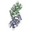
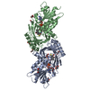
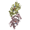
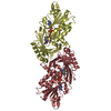
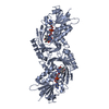
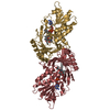
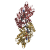
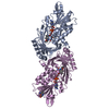
- PDB-4yjf: Crystal structure of DAAO(Y228L/R283G) variant (S-methylbenzylami... -
+ Open data

- Basic information
| Entry | Database: PDB / ID: 4yjf | ||||||
|---|---|---|---|---|---|---|---|
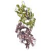
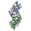
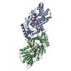
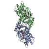
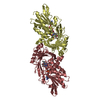
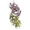
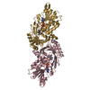
| Title | Crystal structure of DAAO(Y228L/R283G) variant (S-methylbenzylamine binding form) | ||||||
 Components Components | (D-amino-acid ...) x 2 | ||||||
 Keywords Keywords | OXIDOREDUCTASE / amine oxidase / Variant of D-amino acid oxidase / S-methylbenzylamine binding form | ||||||
| Function / homology |  Function and homology information Function and homology informationGlyoxylate metabolism and glycine degradation / Peroxisomal protein import / D-amino-acid oxidase / D-amino-acid oxidase activity / D-alanine catabolic process / glycine oxidase activity / L-proline catabolic process / D-amino acid catabolic process / D-serine catabolic process / presynaptic active zone ...Glyoxylate metabolism and glycine degradation / Peroxisomal protein import / D-amino-acid oxidase / D-amino-acid oxidase activity / D-alanine catabolic process / glycine oxidase activity / L-proline catabolic process / D-amino acid catabolic process / D-serine catabolic process / presynaptic active zone / neutrophil-mediated killing of gram-negative bacterium / dopamine biosynthetic process / peroxisomal matrix / digestion / FAD binding / peroxisome / extracellular region / cytoplasm / cytosol Similarity search - Function | ||||||
| Biological species |  | ||||||
| Method |  X-RAY DIFFRACTION / MOLECULAR REPLACEMENT / Resolution: 2.2 Å X-RAY DIFFRACTION / MOLECULAR REPLACEMENT / Resolution: 2.2 Å | ||||||
 Authors Authors | Nakano, S. / Yasukawa, K. / Kawahara, N. / Ishitsubo, E. / Tokiwa, H. / Asano, Y. | ||||||
 Citation Citation | Journal: To Be Published Title: Crystal structure of DAAO variant Authors: Nakano, S. / Yasukawa, K. / Kawahara, N. / Ishitsubo, E. / Tokiwa, H. / Asano, Y. | ||||||
| History |
|
- Structure visualization
| Structure viewer | Molecule: MolmilJmol/JSmol |
|---|
- Downloads & links
-Download
| PDBx/mmCIF format | 4yjf.cif.gz | 152.9 KB | Display | PDBx/mmCIF format |
|---|---|---|---|---|
| PDB format | pdb4yjf.ent.gz | 118.5 KB | Display | PDB format |
| PDBx/mmJSON format | 4yjf.json.gz | Tree view | PDBx/mmJSON format | |
| Others |  Other downloads Other downloads |
-Validation report
| Arichive directory | https://data.pdbj.org/pub/pdb/validation_reports/yj/4yjfftp://data.pdbj.org/pub/pdb/validation_reports/yj/4yjf | HTTPS FTP |
|---|
-Related structure data
| Related structure data |  4yjdC  4yjgC  4yjhC  3wgtS C: citing same article ( S: Starting model for refinement |
|---|---|
| Similar structure data |


-Links
 PDBj
PDBj- Assembly
| Deposited unit | 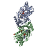
| ||||||||||||||||||
|---|---|---|---|---|---|---|---|---|---|---|---|---|---|---|---|---|---|---|---|
| 1 |
| ||||||||||||||||||
| Unit cell |
| ||||||||||||||||||
| Noncrystallographic symmetry (NCS) | NCS domain:
NCS domain segments: Component-ID: _ / Ens-ID: 1 / Beg auth comp-ID: MET / Beg label comp-ID: MET / End auth comp-ID: ASN / End label comp-ID: ASN / Refine code: _ / Auth seq-ID: 1 - 338 / Label seq-ID: 1 - 338
|
-Components
-D-amino-acid ... , 2 types, 2 molecules AB
| #1: Protein | Mass: 38563.848 Da / Num. of mol.: 1 / Fragment: UNP residues 1-341 / Mutation: Y228L, R283G Source method: isolated from a genetically manipulated source Source: (gene. exp.)  |
|---|---|
| #2: Protein | Mass: 38349.590 Da / Num. of mol.: 1 / Fragment: UNP residues 1-339 / Mutation: Y228L, R283G Source method: isolated from a genetically manipulated source Source: (gene. exp.) |
-Non-polymers , 4 types, 62 molecules 

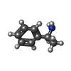

| #3: Chemical |  Mass: 785.550 Da / Num. of mol.: 2 / Source method: obtained synthetically / Formula: C27H33N9O15P2 / Comment: FAD*YM Mass: 785.550 Da / Num. of mol.: 2 / Source method: obtained synthetically / Formula: C27H33N9O15P2 / Comment: FAD*YM#4: Chemical | ChemComp-SO4 /  Mass: 96.063 Da / Num. of mol.: 4 / Source method: obtained synthetically / Formula: SO4 Mass: 96.063 Da / Num. of mol.: 4 / Source method: obtained synthetically / Formula: SO4#5: Chemical |  Mass: 121.180 Da / Num. of mol.: 2 / Source method: obtained synthetically / Formula: C8H11N Mass: 121.180 Da / Num. of mol.: 2 / Source method: obtained synthetically / Formula: C8H11N#6: Water | ChemComp-HOH / | Mass: 18.015 Da / Num. of mol.: 54 / Source method: isolated from a natural source / Formula: H2O |
|---|
-Experimental details
-Experiment
| Experiment | Method: X-RAY DIFFRACTION |
|---|
- Sample preparation
| Crystal | Density Matthews: 2.27 Å3/Da / Density % sol: 45.76 % |
|---|---|
| Crystal grow | Temperature: 293 K / Method: vapor diffusion, sitting drop / pH: 8.5 Details: 30% PEG4000, 0.1M Tris-HCl(pH 8.5), 0.2M Lithium sulfate |
-Data collection
| Diffraction | Mean temperature: 100 K |
|---|---|
| Diffraction source | Source: ROTATING ANODE / Type: RIGAKU MICROMAX-007 / Wavelength: 1.54 Å |
| Detector | Type: RIGAKU RAXIS VII / Detector: IMAGE PLATE / Date: Jun 18, 2013 |
| Radiation | Protocol: SINGLE WAVELENGTH / Monochromatic (M) / Laue (L): M / Scattering type: x-ray |
| Radiation wavelength | Wavelength: 1.54 Å / Relative weight: 1 |
| Reflection | Resolution: 2.2→23.6 Å / Num. obs: 34607 / % possible obs: 95 % / Redundancy: 10.5 % / Rmerge(I) obs: 0.082 / Net I/σ(I): 46.7 |
| Reflection shell | Resolution: 2.2→2.25 Å / Redundancy: 10.2 % / Rmerge(I) obs: 0.41 / Mean I/σ(I) obs: 7.2 / % possible all: 92.3 |
- Processing
| Software |
| ||||||||||||||||||||||||||||||||||||||||||||||||||||||||||||||||||||||||||||||||||||||||||||||||||||||||||||||||||||||||||||||||||||||||||||||||||||||||||||||||||||||||||||||||||||||
|---|---|---|---|---|---|---|---|---|---|---|---|---|---|---|---|---|---|---|---|---|---|---|---|---|---|---|---|---|---|---|---|---|---|---|---|---|---|---|---|---|---|---|---|---|---|---|---|---|---|---|---|---|---|---|---|---|---|---|---|---|---|---|---|---|---|---|---|---|---|---|---|---|---|---|---|---|---|---|---|---|---|---|---|---|---|---|---|---|---|---|---|---|---|---|---|---|---|---|---|---|---|---|---|---|---|---|---|---|---|---|---|---|---|---|---|---|---|---|---|---|---|---|---|---|---|---|---|---|---|---|---|---|---|---|---|---|---|---|---|---|---|---|---|---|---|---|---|---|---|---|---|---|---|---|---|---|---|---|---|---|---|---|---|---|---|---|---|---|---|---|---|---|---|---|---|---|---|---|---|---|---|---|---|
| Refinement | Method to determine structure: MOLECULAR REPLACEMENT Starting model: 3WGT Resolution: 2.2→23.6 Å / Cor.coef. Fo:Fc: 0.953 / Cor.coef. Fo:Fc free: 0.932 / SU B: 5.797 / SU ML: 0.146 / Cross valid method: THROUGHOUT / ESU R: 0.32 / ESU R Free: 0.213 / Stereochemistry target values: MAXIMUM LIKELIHOOD / Details: HYDROGENS HAVE BEEN ADDED IN THE RIDING POSITIONS
| ||||||||||||||||||||||||||||||||||||||||||||||||||||||||||||||||||||||||||||||||||||||||||||||||||||||||||||||||||||||||||||||||||||||||||||||||||||||||||||||||||||||||||||||||||||||
| Solvent computation | Ion probe radii: 0.8 Å / Shrinkage radii: 0.8 Å / VDW probe radii: 1.2 Å / Solvent model: MASK | ||||||||||||||||||||||||||||||||||||||||||||||||||||||||||||||||||||||||||||||||||||||||||||||||||||||||||||||||||||||||||||||||||||||||||||||||||||||||||||||||||||||||||||||||||||||
| Displacement parameters | Biso mean: 33.115 Å2
| ||||||||||||||||||||||||||||||||||||||||||||||||||||||||||||||||||||||||||||||||||||||||||||||||||||||||||||||||||||||||||||||||||||||||||||||||||||||||||||||||||||||||||||||||||||||
| Refinement step | Cycle: 1 / Resolution: 2.2→23.6 Å
| ||||||||||||||||||||||||||||||||||||||||||||||||||||||||||||||||||||||||||||||||||||||||||||||||||||||||||||||||||||||||||||||||||||||||||||||||||||||||||||||||||||||||||||||||||||||
| Refine LS restraints |
|