 Movie
Movie Controller

[English] 日本語
 Yorodumi
- PDB-4yfy: X-ray structure of the Viof N-formyltransferase from Providencia ... -
+ Open data

- Basic information
| Entry | Database: PDB / ID: 4yfy | ||||||
|---|---|---|---|---|---|---|---|
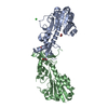
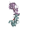
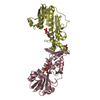
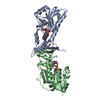
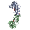
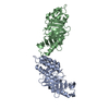
| Title | X-ray structure of the Viof N-formyltransferase from Providencia alcalifaciens O30 in complex with THF and TDP-Qui4N | ||||||
 Components Components | VioF | ||||||
 Keywords Keywords | TRANSFERASE / lipopolysaccharide O-antigen | ||||||
| Function / homology |  Function and homology information Function and homology information | ||||||
| Biological species |  Providencia alcalifaciens (bacteria) Providencia alcalifaciens (bacteria) | ||||||
| Method |  X-RAY DIFFRACTION / MOLECULAR REPLACEMENT / Resolution: 1.9 Å X-RAY DIFFRACTION / MOLECULAR REPLACEMENT / Resolution: 1.9 Å | ||||||
 Authors Authors | Genthe, N.A. / Thoden, J.B. / Benning, M.M. / Holden, H.M. | ||||||
 Citation Citation | Journal: Protein Sci. / Year: 2015 Title: Molecular structure of an N-formyltransferase from Providencia alcalifaciens O30. Authors: Genthe, N.A. / Thoden, J.B. / Benning, M.M. / Holden, H.M. | ||||||
| History |
|
- Structure visualization
| Structure viewer | Molecule: MolmilJmol/JSmol |
|---|
- Downloads & links
-Download
| PDBx/mmCIF format | 4yfy.cif.gz | 127 KB | Display | PDBx/mmCIF format |
|---|---|---|---|---|
| PDB format | pdb4yfy.ent.gz | 97.4 KB | Display | PDB format |
| PDBx/mmJSON format | 4yfy.json.gz | Tree view | PDBx/mmJSON format | |
| Others |  Other downloads Other downloads |
-Validation report
| Summary document | 4yfy_validation.pdf.gz | 1.7 MB | Display | wwPDB validaton report |
|---|---|---|---|---|
| Full document | 4yfy_full_validation.pdf.gz | 1.7 MB | Display | |
| Data in XML | 4yfy_validation.xml.gz | 26.5 KB | Display | |
| Data in CIF | 4yfy_validation.cif.gz | 37.9 KB | Display | |
| Arichive directory | https://data.pdbj.org/pub/pdb/validation_reports/yf/4yfyftp://data.pdbj.org/pub/pdb/validation_reports/yf/4yfy | HTTPS FTP |
-Related structure data
| Related structure data |  4yfvSC S: Starting model for refinement C: citing same article ( |
|---|---|
| Similar structure data |


-Links
 PDBj
PDBj- Assembly
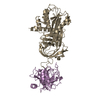
| Deposited unit | 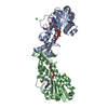
| ||||||||
|---|---|---|---|---|---|---|---|---|---|
| 1 |
| ||||||||
| Unit cell |
| ||||||||
| Components on special symmetry positions |
|
-Components
-Protein , 1 types, 2 molecules AB
| #1: Protein | Mass: 29677.713 Da / Num. of mol.: 2 / Fragment: residues 9-252 Source method: isolated from a genetically manipulated source Source: (gene. exp.) Providencia alcalifaciens (bacteria) / Gene: vioF / Plasmid: pET-DUET / Production host: |
|---|
-Non-polymers , 5 types, 454 molecules 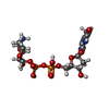


| #2: Chemical |  Mass: 547.345 Da / Num. of mol.: 2 / Source method: obtained synthetically / Formula: C16H27N3O14P2 Mass: 547.345 Da / Num. of mol.: 2 / Source method: obtained synthetically / Formula: C16H27N3O14P2#3: Chemical |  Mass: 445.429 Da / Num. of mol.: 2 / Source method: obtained synthetically / Formula: C19H23N7O6 Mass: 445.429 Da / Num. of mol.: 2 / Source method: obtained synthetically / Formula: C19H23N7O6#4: Chemical | ChemComp-CL / |  Mass: 35.453 Da / Num. of mol.: 1 / Source method: obtained synthetically / Formula: Cl Mass: 35.453 Da / Num. of mol.: 1 / Source method: obtained synthetically / Formula: Cl#5: Chemical | ChemComp-EDO / |  Mass: 62.068 Da / Num. of mol.: 1 / Source method: obtained synthetically / Formula: C2H6O2 Mass: 62.068 Da / Num. of mol.: 1 / Source method: obtained synthetically / Formula: C2H6O2#6: Water | ChemComp-HOH / | Mass: 18.015 Da / Num. of mol.: 448 / Source method: isolated from a natural source / Formula: H2O |
|---|
-Experimental details
-Experiment
| Experiment | Method: X-RAY DIFFRACTION |
|---|
- Sample preparation
| Crystal | Density Matthews: 2.28 Å3/Da / Density % sol: 46.04 % / Description: six-sided stellate rods |
|---|---|
| Crystal grow | Temperature: 277 K / Method: vapor diffusion, hanging drop / pH: 7.5 Details: 8-10% PEG-8000, 1 M tetramethylammonium chloride 100 mM Hepes |
-Data collection
| Diffraction | Mean temperature: 100 K |
|---|---|
| Diffraction source | Source: ROTATING ANODE / Type: RIGAKU RU200 / Wavelength: 1.5418 Å |
| Detector | Type: Bruker Platinum 135 / Detector: CCD / Date: Jun 23, 2014 |
| Radiation | Monochromator: Ni-filter / Protocol: SINGLE WAVELENGTH / Monochromatic (M) / Laue (L): M / Scattering type: x-ray |
| Radiation wavelength | Wavelength: 1.5418 Å / Relative weight: 1 |
| Reflection | Resolution: 1.9→50 Å / Num. all: 42792 / Num. obs: 42792 / % possible obs: 99.9 % / Observed criterion σ(F): 0 / Observed criterion σ(I): 0 / Redundancy: 5.3 % / Rmerge(I) obs: 0.053 / Rsym value: 0.053 / Net I/av σ(I): 14.3 / Net I/σ(I): 14.3 |
| Reflection shell | Resolution: 1.9→2 Å / Redundancy: 4.5 % / Rmerge(I) obs: 0.237 / Mean I/σ(I) obs: 2.4 / % possible all: 99.1 |
- Processing
| Software |
| ||||||||||||||||||||||||||||||||||||||||||||||||||||||||||||||||||||||||||||||||||||||||||||||||||||||||||||||||||||||||||||||||||||||||||||||||||||||||||||||||||||||||||||||||||||||
|---|---|---|---|---|---|---|---|---|---|---|---|---|---|---|---|---|---|---|---|---|---|---|---|---|---|---|---|---|---|---|---|---|---|---|---|---|---|---|---|---|---|---|---|---|---|---|---|---|---|---|---|---|---|---|---|---|---|---|---|---|---|---|---|---|---|---|---|---|---|---|---|---|---|---|---|---|---|---|---|---|---|---|---|---|---|---|---|---|---|---|---|---|---|---|---|---|---|---|---|---|---|---|---|---|---|---|---|---|---|---|---|---|---|---|---|---|---|---|---|---|---|---|---|---|---|---|---|---|---|---|---|---|---|---|---|---|---|---|---|---|---|---|---|---|---|---|---|---|---|---|---|---|---|---|---|---|---|---|---|---|---|---|---|---|---|---|---|---|---|---|---|---|---|---|---|---|---|---|---|---|---|---|---|
| Refinement | Method to determine structure: MOLECULAR REPLACEMENT Starting model: 4YFV Resolution: 1.9→50 Å / Cor.coef. Fo:Fc: 0.952 / Cor.coef. Fo:Fc free: 0.912 / SU B: 3.978 / SU ML: 0.116 / Cross valid method: THROUGHOUT / ESU R: 0.162 / ESU R Free: 0.158 / Stereochemistry target values: MAXIMUM LIKELIHOOD / Details: HYDROGENS HAVE BEEN ADDED IN THE RIDING POSITIONS
| ||||||||||||||||||||||||||||||||||||||||||||||||||||||||||||||||||||||||||||||||||||||||||||||||||||||||||||||||||||||||||||||||||||||||||||||||||||||||||||||||||||||||||||||||||||||
| Solvent computation | Ion probe radii: 0.8 Å / Shrinkage radii: 0.8 Å / VDW probe radii: 1.2 Å / Solvent model: MASK | ||||||||||||||||||||||||||||||||||||||||||||||||||||||||||||||||||||||||||||||||||||||||||||||||||||||||||||||||||||||||||||||||||||||||||||||||||||||||||||||||||||||||||||||||||||||
| Displacement parameters | Biso mean: 22.31 Å2
| ||||||||||||||||||||||||||||||||||||||||||||||||||||||||||||||||||||||||||||||||||||||||||||||||||||||||||||||||||||||||||||||||||||||||||||||||||||||||||||||||||||||||||||||||||||||
| Refinement step | Cycle: 1 / Resolution: 1.9→50 Å
| ||||||||||||||||||||||||||||||||||||||||||||||||||||||||||||||||||||||||||||||||||||||||||||||||||||||||||||||||||||||||||||||||||||||||||||||||||||||||||||||||||||||||||||||||||||||
| Refine LS restraints |
|