 Movie
Movie Controller

[English] 日本語
 Yorodumi
- PDB-4yca: Evidence of Kinetic Cooperativity in dimeric Ketopantoate Reducta... -
+ Open data

- Basic information
| Entry | Database: PDB / ID: 4yca | ||||||
|---|---|---|---|---|---|---|---|
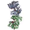
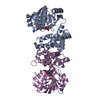
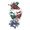
| Title | Evidence of Kinetic Cooperativity in dimeric Ketopantoate Reductase from Staphylococcus aureus | ||||||
 Components Components | 2-dehydropantoate 2-reductase | ||||||
 Keywords Keywords | OXIDOREDUCTASE / Reductase / Dimer / Rossmann / kinetic cooperativity | ||||||
| Function / homology |  Function and homology information Function and homology information2-dehydropantoate 2-reductase / 2-dehydropantoate 2-reductase activity / phosphogluconate dehydrogenase (decarboxylating) activity / pantothenate biosynthetic process / nucleotide binding / cytoplasm Similarity search - Function | ||||||
| Biological species |   Staphylococcus aureus (bacteria) Staphylococcus aureus (bacteria) | ||||||
| Method |  X-RAY DIFFRACTION / SYNCHROTRON / MOLECULAR REPLACEMENT / Resolution: 1.81 Å X-RAY DIFFRACTION / SYNCHROTRON / MOLECULAR REPLACEMENT / Resolution: 1.81 Å | ||||||
 Authors Authors | Sanchez, J.E. / Gross, P.G. / Goetze, R. / Walsh Jr., R.M. / Peeples, W.B. / Wood, Z.A. | ||||||
 Citation Citation | Journal: Biochemistry / Year: 2015 Title: Evidence of Kinetic Cooperativity in Dimeric Ketopantoate Reductase from Staphylococcus aureus. Authors: Sanchez, J.E. / Gross, P.G. / Goetze, R.W. / Walsh Jr., R.M. / Peeples, W.B. / Wood, Z.A. | ||||||
| History |
|
- Structure visualization
| Structure viewer | Molecule: MolmilJmol/JSmol |
|---|
- Downloads & links
-Download
| PDBx/mmCIF format | 4yca.cif.gz | 252.7 KB | Display | PDBx/mmCIF format |
|---|---|---|---|---|
| PDB format | pdb4yca.ent.gz | 203.3 KB | Display | PDB format |
| PDBx/mmJSON format | 4yca.json.gz | Tree view | PDBx/mmJSON format | |
| Others |  Other downloads Other downloads |
-Validation report
| Arichive directory | https://data.pdbj.org/pub/pdb/validation_reports/yc/4ycaftp://data.pdbj.org/pub/pdb/validation_reports/yc/4yca | HTTPS FTP |
|---|
-Related structure data
| Related structure data |  4s3mC 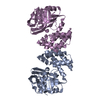 3g17S C: citing same article ( S: Starting model for refinement |
|---|---|
| Similar structure data |


-Links
 PDBj
PDBj


- Assembly
| Deposited unit | 
| ||||||||
|---|---|---|---|---|---|---|---|---|---|
| 1 |
| ||||||||
| Unit cell |
|
-Components
| #1: Protein | Mass: 33435.938 Da / Num. of mol.: 2 Source method: isolated from a genetically manipulated source Source: (gene. exp.) Staphylococcus aureus (bacteria) / Gene: CH52_06005, DA92_02185 / Production host: References: UniProt: W8TQG0, UniProt: A0A0J9X283*PLUS, 2-dehydropantoate 2-reductase #2: Chemical |   Mass: 745.421 Da / Num. of mol.: 2 / Source method: obtained synthetically / Formula: C21H30N7O17P3 Mass: 745.421 Da / Num. of mol.: 2 / Source method: obtained synthetically / Formula: C21H30N7O17P3#3: Water | ChemComp-HOH / |  Mass: 18.015 Da / Num. of mol.: 411 / Source method: isolated from a natural source / Formula: H2O Mass: 18.015 Da / Num. of mol.: 411 / Source method: isolated from a natural source / Formula: H2O |
|---|
-Experimental details
-Experiment
| Experiment | Method: X-RAY DIFFRACTION |
|---|
- Sample preparation
| Crystal | Density Matthews: 2.38 Å3/Da / Density % sol: 48.3 % |
|---|---|
| Crystal grow | Temperature: 293 K / Method: vapor diffusion, sitting drop / pH: 6.6 Details: 4mM KP, 4mM NADP+, 300mM Magnesium acetate, 100mM MES, 15% PEG 3,350 |
-Data collection
| Diffraction | Mean temperature: 77 K |
|---|---|
| Diffraction source | Source: SYNCHROTRON / Site: APS  / Beamline: 22-BM / Wavelength: 1 Å / Beamline: 22-BM / Wavelength: 1 Å |
| Detector | Type: MAR scanner 300 mm plate / Detector: IMAGE PLATE / Date: Jun 19, 2013 |
| Radiation | Protocol: SINGLE WAVELENGTH / Monochromatic (M) / Laue (L): M / Scattering type: x-ray |
| Radiation wavelength | Wavelength: 1 Å / Relative weight: 1 |
| Reflection | Resolution: 1.81→44.3 Å / Num. obs: 59348 / % possible obs: 99.9 % / Redundancy: 13.8 % / Net I/σ(I): 1.34 |
- Processing
| Software |
| ||||||||||||||||||||||||||||||||||||||||||||||||||||||||||||||||||||||||||||||||||||||||||||||||||||||||||||||||
|---|---|---|---|---|---|---|---|---|---|---|---|---|---|---|---|---|---|---|---|---|---|---|---|---|---|---|---|---|---|---|---|---|---|---|---|---|---|---|---|---|---|---|---|---|---|---|---|---|---|---|---|---|---|---|---|---|---|---|---|---|---|---|---|---|---|---|---|---|---|---|---|---|---|---|---|---|---|---|---|---|---|---|---|---|---|---|---|---|---|---|---|---|---|---|---|---|---|---|---|---|---|---|---|---|---|---|---|---|---|---|---|---|---|
| Refinement | Method to determine structure: MOLECULAR REPLACEMENT Starting model: 3G17 Resolution: 1.81→44.3 Å / SU ML: 0.19 / Cross valid method: FREE R-VALUE / σ(F): 1.34 / Phase error: 20.95 / Stereochemistry target values: ML
| ||||||||||||||||||||||||||||||||||||||||||||||||||||||||||||||||||||||||||||||||||||||||||||||||||||||||||||||||
| Solvent computation | Shrinkage radii: 0.9 Å / VDW probe radii: 1.11 Å / Solvent model: FLAT BULK SOLVENT MODEL | ||||||||||||||||||||||||||||||||||||||||||||||||||||||||||||||||||||||||||||||||||||||||||||||||||||||||||||||||
| Refinement step | Cycle: LAST / Resolution: 1.81→44.3 Å
| ||||||||||||||||||||||||||||||||||||||||||||||||||||||||||||||||||||||||||||||||||||||||||||||||||||||||||||||||
| Refine LS restraints |
| ||||||||||||||||||||||||||||||||||||||||||||||||||||||||||||||||||||||||||||||||||||||||||||||||||||||||||||||||
| LS refinement shell |
|
