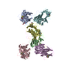
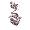
| Deposited unit | A: Coat protein
B: Coat protein
C: Coat protein
D: Coat protein
E: Coat protein
F: Coat protein
hetero molecules
| Theoretical mass | Number of molelcules |
|---|
| Total (without water) | 111,719 | 7 |
|---|
| Polymers | 111,657 | 6 |
|---|
| Non-polymers | 62 | 1 |
|---|
| Water | 7,692 | 427 |
|---|
|
|---|
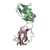
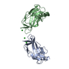
| 1 | A: Coat protein
F: Coat protein
hetero molecules
| Theoretical mass | Number of molelcules |
|---|
| Total (without water) | 37,281 | 3 |
|---|
| Polymers | 37,219 | 2 |
|---|
| Non-polymers | 62 | 1 |
|---|
| Water | 36 | 2 |
|---|
| Type | Name | Symmetry operation | Number |
|---|
| identity operation | 1_555 | x,y,z | 1 |
| Buried area | 2840 Å2 |
|---|
| ΔGint | -14 kcal/mol |
|---|
| Surface area | 15160 Å2 |
|---|
| Method | PISA |
|---|
|
|---|
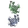
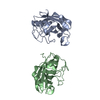
| 2 | B: Coat protein
C: Coat protein
| Theoretical mass | Number of molelcules |
|---|
| Total (without water) | 37,219 | 2 |
|---|
| Polymers | 37,219 | 2 |
|---|
| Non-polymers | 0 | 0 |
|---|
| Water | 36 | 2 |
|---|
| Type | Name | Symmetry operation | Number |
|---|
| identity operation | 1_555 | x,y,z | 1 |
| Buried area | 2590 Å2 |
|---|
| ΔGint | -19 kcal/mol |
|---|
| Surface area | 15160 Å2 |
|---|
| Method | PISA |
|---|
|
|---|
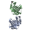
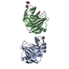
| 3 | D: Coat protein
E: Coat protein
| Theoretical mass | Number of molelcules |
|---|
| Total (without water) | 37,219 | 2 |
|---|
| Polymers | 37,219 | 2 |
|---|
| Non-polymers | 0 | 0 |
|---|
| Water | 36 | 2 |
|---|
| Type | Name | Symmetry operation | Number |
|---|
| identity operation | 1_555 | x,y,z | 1 |
| Buried area | 2570 Å2 |
|---|
| ΔGint | -18 kcal/mol |
|---|
| Surface area | 15560 Å2 |
|---|
| Method | PISA |
|---|
|
|---|
| Unit cell | | Length a, b, c (Å) | 103.040, 69.980, 103.440 |
|---|
| Angle α, β, γ (deg.) | 90.000, 105.510, 90.000 |
|---|
| Int Tables number | 4 |
|---|
| Space group name H-M | P1211 |
|---|
|
|---|
| Noncrystallographic symmetry (NCS) | NCS domain: | ID | Ens-ID | Details (eV) |
|---|
| 1 | 1 | A| 2 | 1 | B| 1 | 2 | A| 2 | 2 | C| 1 | 3 | A| 2 | 3 | D| 1 | 4 | A| 2 | 4 | E| 1 | 5 | A| 2 | 5 | F| 1 | 6 | B| 2 | 6 | C| 1 | 7 | B| 2 | 7 | D| 1 | 8 | B| 2 | 8 | E| 1 | 9 | B| 2 | 9 | F| 1 | 10 | C| 2 | 10 | D| 1 | 11 | C| 2 | 11 | E| 1 | 12 | C| 2 | 12 | F| 1 | 13 | D| 2 | 13 | E| 1 | 14 | D| 2 | 14 | F| 1 | 15 | E| 2 | 15 | F | | | | | | | | | | | | | | | | | | | | | | | | | | | | | |
NCS domain segments: Component-ID: _ / End auth comp-ID: ASP / End label comp-ID: ASP / Refine code: _ | Dom-ID | Ens-ID | Beg auth comp-ID | Beg label comp-ID | Auth asym-ID | Label asym-ID | Auth seq-ID | Label seq-ID |
|---|
| 1 | 1 | SERSERAA| 90 - 238 | 18 - 166 | | 2 | 1 | SERSERBB| 90 - 238 | 18 - 166 | | 1 | 2 | LEULEUAA| 91 - 238 | 19 - 166 | | 2 | 2 | LEULEUCC| 91 - 238 | 19 - 166 | | 1 | 3 | LEULEUAA| 91 - 238 | 19 - 166 | | 2 | 3 | LEULEUDD| 91 - 238 | 19 - 166 | | 1 | 4 | SERSERAA| 90 - 238 | 18 - 166 | | 2 | 4 | SERSEREE| 90 - 238 | 18 - 166 | | 1 | 5 | SERSERAA| 90 - 238 | 18 - 166 | | 2 | 5 | SERSERFF| 90 - 238 | 18 - 166 | | 1 | 6 | LEULEUBB| 91 - 238 | 19 - 166 | | 2 | 6 | LEULEUCC| 91 - 238 | 19 - 166 | | 1 | 7 | LEULEUBB| 91 - 238 | 19 - 166 | | 2 | 7 | LEULEUDD| 91 - 238 | 19 - 166 | | 1 | 8 | SERSERBB| 90 - 238 | 18 - 166 | | 2 | 8 | SERSEREE| 90 - 238 | 18 - 166 | | 1 | 9 | SERSERBB| 90 - 238 | 18 - 166 | | 2 | 9 | SERSERFF| 90 - 238 | 18 - 166 | | 1 | 10 | LEULEUC| C | | | | | | | | | | | | | | | | | | | | | | | | | | | | | | | | | | | | | | | | | | | | | | | | | | | | | | | | | | | | | | | | | | | | | | | | | | | |
|
|---|
 Movie
Movie Controller

 Yorodumi
 Open data
 Basic information
 Components
 Keywords
 Function and homology information
 Tobacco streak virus
 X-RAY DIFFRACTION /
  Authors
 India, 1items
  Citation
 Structure visualization
 Downloads & links
 Other downloads


 PDBj
PDBj Assembly