 Movie
Movie Controller

[English] 日本語
 Yorodumi
- PDB-4xwa: TMK from S.aureus in complex with the Piperidinyl Thymine class i... -
+ Open data

- Basic information
| Entry | Database: PDB / ID: 4xwa | ||||||
|---|---|---|---|---|---|---|---|
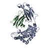
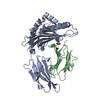
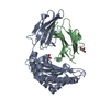
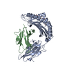
| Title | TMK from S.aureus in complex with the Piperidinyl Thymine class inhibitor with a C5 ethyl-amine | ||||||
 Components Components | Thymidylate kinase | ||||||
 Keywords Keywords | TRANSFERASE / TMK / kinase / antibacterial / piperidine / thymidine | ||||||
| Function / homology |  Function and homology information Function and homology informationdTMP kinase / dUDP biosynthetic process / dTDP biosynthetic process / dTMP kinase activity / dTTP biosynthetic process / ATP binding / cytosol Similarity search - Function | ||||||
| Biological species |   Staphylococcus aureus (bacteria) Staphylococcus aureus (bacteria) | ||||||
| Method |  X-RAY DIFFRACTION / MOLECULAR REPLACEMENT / molecular replacement / Resolution: 1.89 Å X-RAY DIFFRACTION / MOLECULAR REPLACEMENT / molecular replacement / Resolution: 1.89 Å | ||||||
 Authors Authors | Olivier, N.B. | ||||||
 Citation Citation | Journal: Bioorg Med Chem Lett / Year: 2015 Title: A highly potent antibacterial inhibitor of Gram-positive bacterial thymidylate kinase (TMK): SAR of piperidinyl thymines at position C5 and L1 Authors: Guler, S.Y. / Martinez-Botella, G. / Breen, J. / Kawatkar, S. / Loch, J. / Olivier, N.B. / Wang, H. / Keating, T. / Otterson, L. | ||||||
| History |
|
- Structure visualization
| Structure viewer | Molecule: MolmilJmol/JSmol |
|---|
- Downloads & links
-Download
| PDBx/mmCIF format | 4xwa.cif.gz | 101.1 KB | Display | PDBx/mmCIF format |
|---|---|---|---|---|
| PDB format | pdb4xwa.ent.gz | 76.1 KB | Display | PDB format |
| PDBx/mmJSON format | 4xwa.json.gz | Tree view | PDBx/mmJSON format | |
| Others |  Other downloads Other downloads |
-Validation report
| Arichive directory | https://data.pdbj.org/pub/pdb/validation_reports/xw/4xwaftp://data.pdbj.org/pub/pdb/validation_reports/xw/4xwa | HTTPS FTP |
|---|
-Related structure data
| Similar structure data |
|---|


-Links
 PDBj
PDBj- Assembly
| Deposited unit | 
| ||||||||
|---|---|---|---|---|---|---|---|---|---|
| 1 |
| ||||||||
| Unit cell |
| ||||||||
| Details | biological unit is the same as asym. |
-Components
| #1: Protein | Mass: 23454.586 Da / Num. of mol.: 2 Source method: isolated from a genetically manipulated source Source: (gene. exp.) Staphylococcus aureus (strain Mu3 / ATCC 700698) (bacteria)Gene: tmk, SAHV_0479 / Production host: #2: Chemical |   Mass: 589.081 Da / Num. of mol.: 2 / Source method: obtained synthetically / Formula: C32H33ClN4O5 Mass: 589.081 Da / Num. of mol.: 2 / Source method: obtained synthetically / Formula: C32H33ClN4O5#3: Water | ChemComp-HOH / |  Mass: 18.015 Da / Num. of mol.: 311 / Source method: isolated from a natural source / Formula: H2O Mass: 18.015 Da / Num. of mol.: 311 / Source method: isolated from a natural source / Formula: H2O |
|---|
-Experimental details
-Experiment
| Experiment | Method: X-RAY DIFFRACTION / Number of used crystals: 1 |
|---|
- Sample preparation
| Crystal | Density Matthews: 2.19 Å3/Da / Density % sol: 43.73 % / Description: bars |
|---|---|
| Crystal grow | Temperature: 293 K / Method: vapor diffusion, sitting drop / pH: 7 Details: To obtain the inhibitor bound crystal form of TMK-S.aureus crystals were initially grown in the absence of compound using the sitting drop method at 293 K with a reservoir solution of 100 mM ...Details: To obtain the inhibitor bound crystal form of TMK-S.aureus crystals were initially grown in the absence of compound using the sitting drop method at 293 K with a reservoir solution of 100 mM PCPT (propionate-cacodylate-bistris propane buffer) pH 7-8, 21-24% PEG 3350, 200 mM Mg2Cl using 1:1 protein:reservoir solution with the protein solution at 13 mg/mL. Crystals were harvested and soaked overnight in a solution containing 100 mM PCPT, 35% PEG 3350, 200 mM Mg2Cl and 1-2 mM TK-666 from a 100 mM DMSO stock. After soaking the crystals were cryoprotected by soaking for 15 minutes in compound-soak solution supplemented with 20% ethylene glycol. Cryoprotected crystals were mounted on nylon loops and flash-cooled in liquid nitrogen. PH range: 7-8 |
-Data collection
| Diffraction | Mean temperature: 100 K | ||||||||||||||||||||||||||||||||||||||||||||||||||||||||||||||||||||||||||||||||||||||||||||||||||||||||||||||
|---|---|---|---|---|---|---|---|---|---|---|---|---|---|---|---|---|---|---|---|---|---|---|---|---|---|---|---|---|---|---|---|---|---|---|---|---|---|---|---|---|---|---|---|---|---|---|---|---|---|---|---|---|---|---|---|---|---|---|---|---|---|---|---|---|---|---|---|---|---|---|---|---|---|---|---|---|---|---|---|---|---|---|---|---|---|---|---|---|---|---|---|---|---|---|---|---|---|---|---|---|---|---|---|---|---|---|---|---|---|---|---|
| Diffraction source | Source: ROTATING ANODE / Type: RIGAKU FR-E+ SUPERBRIGHT / Wavelength: 1.54 Å | ||||||||||||||||||||||||||||||||||||||||||||||||||||||||||||||||||||||||||||||||||||||||||||||||||||||||||||||
| Detector | Type: RIGAKU SATURN 944+ / Detector: CCD / Date: Jun 6, 2011 | ||||||||||||||||||||||||||||||||||||||||||||||||||||||||||||||||||||||||||||||||||||||||||||||||||||||||||||||
| Radiation | Protocol: SINGLE WAVELENGTH / Monochromatic (M) / Laue (L): M / Scattering type: x-ray | ||||||||||||||||||||||||||||||||||||||||||||||||||||||||||||||||||||||||||||||||||||||||||||||||||||||||||||||
| Radiation wavelength | Wavelength: 1.54 Å / Relative weight: 1 | ||||||||||||||||||||||||||||||||||||||||||||||||||||||||||||||||||||||||||||||||||||||||||||||||||||||||||||||
| Reflection | Resolution: 1.887→33.831 Å / Num. all: 31072 / Num. obs: 31072 / % possible obs: 95.9 % / Redundancy: 2.8 % / Biso Wilson estimate: 23.81 Å2 / Rpim(I) all: 0.029 / Rrim(I) all: 0.053 / Rsym value: 0.037 / Net I/av σ(I): 15.813 / Net I/σ(I): 16.2 / Num. measured all: 88375 | ||||||||||||||||||||||||||||||||||||||||||||||||||||||||||||||||||||||||||||||||||||||||||||||||||||||||||||||
| Reflection shell | Diffraction-ID: 1 / Rejects: _
|
-Phasing
| Phasing | Method: molecular replacement |
|---|
- Processing
| Software |
| ||||||||||||||||||||||||||||||||||||||||||||||||||||||||||||||||||||||||||||||||||||||||||||||||||||||||||||
|---|---|---|---|---|---|---|---|---|---|---|---|---|---|---|---|---|---|---|---|---|---|---|---|---|---|---|---|---|---|---|---|---|---|---|---|---|---|---|---|---|---|---|---|---|---|---|---|---|---|---|---|---|---|---|---|---|---|---|---|---|---|---|---|---|---|---|---|---|---|---|---|---|---|---|---|---|---|---|---|---|---|---|---|---|---|---|---|---|---|---|---|---|---|---|---|---|---|---|---|---|---|---|---|---|---|---|---|---|---|
| Refinement | Method to determine structure: MOLECULAR REPLACEMENT Starting model: apo-state structure Resolution: 1.89→33.83 Å / Cor.coef. Fo:Fc: 0.94 / Cor.coef. Fo:Fc free: 0.9136 / SU R Cruickshank DPI: 0.169 / Cross valid method: THROUGHOUT / σ(F): 0 / SU R Blow DPI: 0.173 / SU Rfree Blow DPI: 0.149 / SU Rfree Cruickshank DPI: 0.148
| ||||||||||||||||||||||||||||||||||||||||||||||||||||||||||||||||||||||||||||||||||||||||||||||||||||||||||||
| Displacement parameters | Biso max: 114.56 Å2 / Biso mean: 27.39 Å2 / Biso min: 9.27 Å2
| ||||||||||||||||||||||||||||||||||||||||||||||||||||||||||||||||||||||||||||||||||||||||||||||||||||||||||||
| Refine analyze | Luzzati coordinate error obs: 0.228 Å | ||||||||||||||||||||||||||||||||||||||||||||||||||||||||||||||||||||||||||||||||||||||||||||||||||||||||||||
| Refinement step | Cycle: final / Resolution: 1.89→33.83 Å
| ||||||||||||||||||||||||||||||||||||||||||||||||||||||||||||||||||||||||||||||||||||||||||||||||||||||||||||
| Refine LS restraints |
| ||||||||||||||||||||||||||||||||||||||||||||||||||||||||||||||||||||||||||||||||||||||||||||||||||||||||||||
| LS refinement shell | Resolution: 1.89→1.95 Å / Total num. of bins used: 16
|
