Monochromator: Cu / Protocol: SINGLE WAVELENGTH / Monochromatic (M) / Laue (L): M / Scattering type: x-ray
Radiation wavelength
Wavelength: 1.54 Å / Relative weight: 1
Reflection
Resolution: 2→27 Å / Num. obs: 39530 / % possible obs: 99.3 % / Redundancy: 3.6 % / Net I/σ(I): 11.64
-
Processing
Software
Name
Version
Classification
REFMAC
5.7.0032
refinement
HKL-2000
datareduction
DENZO
datascaling
MOLREP
modelbuilding
Refinement
Resolution: 2→23.47 Å / Cor.coef. Fo:Fc: 0.954 / Cor.coef. Fo:Fc free: 0.925 / SU B: 4.257 / SU ML: 0.12 / Cross valid method: THROUGHOUT / ESU R: 0.202 / ESU R Free: 0.179 / Stereochemistry target values: MAXIMUM LIKELIHOOD / Details: HYDROGENS HAVE BEEN ADDED IN THE RIDING POSITIONS
Rfactor
Num. reflection
% reflection
Selection details
Rfree
0.2312
1979
5 %
RANDOM
Rwork
0.17607
-
-
-
obs
0.17895
37550
92.44 %
-
Solvent computation
Ion probe radii: 0.8 Å / Shrinkage radii: 0.8 Å / VDW probe radii: 1.2 Å / Solvent model: MASK
 Movie
Movie Controller

 Yorodumi
 Open data
 Basic information
 Components
 Keywords
 Function and homology information
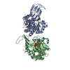
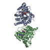
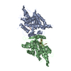
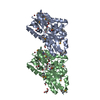
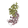
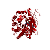
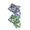
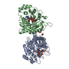
 Mycobacterium tuberculosis H37Ra (bacteria)
 X-RAY DIFFRACTION / Resolution: 2 Å
  Authors
 Citation
 Structure visualization
 Downloads & links
 Other downloads
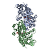

 PDBj
PDBj Assembly
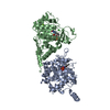

 Mass: 456.344 Da / Num. of mol.: 2 / Source method: obtained synthetically / Formula: C17H21N4O9P
 Mass: 35.453 Da / Num. of mol.: 5 / Source method: obtained synthetically / Formula: Cl
 Mass: 24.305 Da / Num. of mol.: 1 / Source method: obtained synthetically / Formula: Mg
 Mass: 18.015 Da / Num. of mol.: 213 / Source method: isolated from a natural source / Formula: H2O
 Sample preparation
 Processing