Component-ID: 1 / Ens-ID: 1 / Beg auth comp-ID: SER / Beg label comp-ID: SER / End auth comp-ID: LEU / End label comp-ID: LEU / Auth seq-ID: 3 - 401 / Label seq-ID: 19 - 417
Dom-ID
Selection details
Auth asym-ID
Label asym-ID
1
chainA
A
A
2
chainB
B
B
3
chainC
C
C
4
chainD
D
D
5
chainE
E
E
6
chainF
F
F
7
chainG
G
G
8
chainH
H
H
9
chainI
I
I
10
chainJ
J
J
11
chainK
K
K
12
chainL
L
L
13
chainM
M
M
-
Components
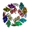
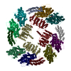
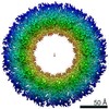
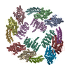
#1: Protein
Nucleocapsid / Nucleocapsid protein
Mass: 58399.004 Da / Num. of mol.: 13 Source method: isolated from a genetically manipulated source Source: (gene. exp.) Parainfluenza virus 5 / Production host: Escherichia coli (E. coli) / References: UniProt: W5QKM4, UniProt: Q88435*PLUS
#2: RNA chain
RNA (78-MER)
Mass: 23835.986 Da / Num. of mol.: 1 Source method: isolated from a genetically manipulated source Source: (gene. exp.) Escherichia coli (E. coli) / Production host: Escherichia coli (E. coli)
Mass: 207.200 Da / Num. of mol.: 14 / Source method: obtained synthetically / Formula: Pb
Has protein modification
Y
-
Experimental details
-
Experiment
Experiment
Method: X-RAY DIFFRACTION / Number of used crystals: 1
-
Sample preparation
Crystal
Density Matthews: 3.07 Å3/Da / Density % sol: 59.95 %
Crystal grow
Temperature: 298 K / Method: vapor diffusion, sitting drop / pH: 6.5 Details: Crystlliztion Condition: 30% PEG 400, 0.1M MES sodium salt pH 6.5, 0.1 M MgCl2. Crystals grew from 4 days to one week, and varied in size from 50um to 400um PH range: 6.5-7.2 / Temp details: room temperature
In the structure databanks used in Yorodumi, some data are registered as the other names, "COVID-19 virus" and "2019-nCoV". Here are the details of the virus and the list of structure data.
Jan 31, 2019. EMDB accession codes are about to change! (news from PDBe EMDB page)
EMDB accession codes are about to change! (news from PDBe EMDB page)
The allocation of 4 digits for EMDB accession codes will soon come to an end. Whilst these codes will remain in use, new EMDB accession codes will include an additional digit and will expand incrementally as the available range of codes is exhausted. The current 4-digit format prefixed with “EMD-” (i.e. EMD-XXXX) will advance to a 5-digit format (i.e. EMD-XXXXX), and so on. It is currently estimated that the 4-digit codes will be depleted around Spring 2019, at which point the 5-digit format will come into force.
The EM Navigator/Yorodumi systems omit the EMD- prefix.
Related info.:Q: What is EMD? / ID/Accession-code notation in Yorodumi/EM Navigator
Yorodumi is a browser for structure data from EMDB, PDB, SASBDB, etc.
This page is also the successor to EM Navigator detail page, and also detail information page/front-end page for Omokage search.
The word "yorodu" (or yorozu) is an old Japanese word meaning "ten thousand". "mi" (miru) is to see.
Related info.:EMDB / PDB / SASBDB / Comparison of 3 databanks / Yorodumi Search / Aug 31, 2016. New EM Navigator & Yorodumi / Yorodumi Papers / Jmol/JSmol / Function and homology information / Changes in new EM Navigator and Yorodumi
 Movie
Movie Controller

 Yorodumi
 Open data
 Basic information
 Components
 Keywords
 Function and homology information
 Parainfluenza virus 5
 X-RAY DIFFRACTION /
  Authors
 Citation
 Structure visualization
 Downloads & links
 Other downloads
 PDBj
PDBj


 Assembly
 Mass: 207.200 Da / Num. of mol.: 14 / Source method: obtained synthetically / Formula: Pb
 Sample preparation
 / Beamline: 21-ID-D / Wavelength: 0.95041 Å
 Processing