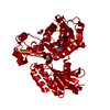
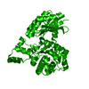
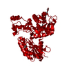
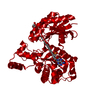
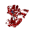
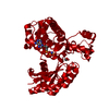
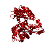
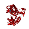
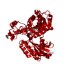
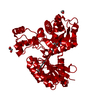
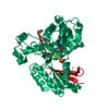
Entry Database : PDB / ID : 4xh5Title Crystal structure of Salmonella typhimurium propionate kinase A88G mutant, in complex with AMPPNP and propionate Propionate kinase Keywords / / / / Function / homology Function Domain/homology Component
/ / / / / / / / / / / / / / / / / / / / / / / / / / / Biological species Salmonella typhimurium (bacteria)Method / / / Resolution : 2.11 Å Authors Murthy, A.M. / Mathivanan, S. / Chittori, S. / Savithri, H.S. / Murthy, M.R.N. Funding support Organization Grant number Country DBT
Journal : Acta Crystallogr.,Sect.D / Year : 2015Title : Structures of substrate- and nucleotide-bound propionate kinase from Salmonella typhimurium: substrate specificity and phosphate-transfer mechanismAuthors : Murthy, A.M.V. / Mathivanan, S. / Chittori, S. / Savithri, H.S. / Murthy, M.R.N. History Deposition Jan 4, 2015 Deposition site / Processing site Revision 1.0 Sep 23, 2015 Provider / Type Revision 1.1 Nov 8, 2023 Group Data collection / Database references ... Data collection / Database references / Derived calculations / Refinement description Category chem_comp_atom / chem_comp_bond ... chem_comp_atom / chem_comp_bond / database_2 / pdbx_initial_refinement_model / pdbx_struct_oper_list Item / _database_2.pdbx_database_accession / _pdbx_struct_oper_list.symmetry_operation
Show all Show less
 Movie
Movie Controller

 Yorodumi
 Open data
 Basic information
 Components
 Keywords
 Function and homology information
 Salmonella typhimurium (bacteria)
 X-RAY DIFFRACTION /
  Authors
 India, 1items
  Citation
 Structure visualization
 Downloads & links
 Other downloads
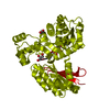


 PDBj
PDBj


 Assembly
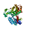


 Mass: 96.063 Da / Num. of mol.: 1 / Source method: obtained synthetically / Formula: SO4
 Mass: 92.094 Da / Num. of mol.: 1 / Source method: obtained synthetically / Formula: C3H8O3
 Mass: 506.196 Da / Num. of mol.: 1 / Source method: obtained synthetically / Formula: C10H17N6O12P3 / Comment: AMP-PNP, energy-carrying molecule analogue*YM
 Mass: 74.079 Da / Num. of mol.: 1 / Source method: obtained synthetically / Formula: C3H6O2
 Sample preparation
 / Beamline: BM14 / Wavelength: 0.976 Å
 Processing