 Movie
Movie Controller

[English] 日本語
 Yorodumi
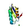
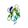
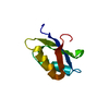
- PDB-4xfn: Structure of an Amyloid forming peptide AEVVFT from Human Transth... -
+ Open data

- Basic information
| Entry | Database: PDB / ID: 4xfn | ||||||
|---|---|---|---|---|---|---|---|
| Title | Structure of an Amyloid forming peptide AEVVFT from Human Transthyretin | ||||||
 Components Components | Amyloid forming peptide AEVVFT | ||||||
 Keywords Keywords | PROTEIN FIBRIL / amyloid / transthyretin / fibril | ||||||
| Biological species | synthetic construct (others) | ||||||
| Method |  X-RAY DIFFRACTION / SYNCHROTRON / MOLECULAR REPLACEMENT / Resolution: 1.85 Å X-RAY DIFFRACTION / SYNCHROTRON / MOLECULAR REPLACEMENT / Resolution: 1.85 Å | ||||||
 Authors Authors | Saelices, L. / Sawaya, M. / Cascio, D. / Eisenberg, D.S. | ||||||
| Funding support |  Switzerland, 1items Switzerland, 1items
| ||||||
 Citation Citation | Journal: J.Biol.Chem. / Year: 2015 Title: Uncovering the Mechanism of Aggregation of Human Transthyretin. Authors: Saelices, L. / Johnson, L.M. / Liang, W.Y. / Sawaya, M.R. / Cascio, D. / Ruchala, P. / Whitelegge, J. / Jiang, L. / Riek, R. / Eisenberg, D.S. | ||||||
| History |
|
- Structure visualization
| Structure viewer | Molecule:  MolmilJmol/JSmol MolmilJmol/JSmol |
|---|
- Downloads & links
-Download
| PDBx/mmCIF format | 4xfn.cif.gz | 10.1 KB | Display | PDBx/mmCIF format |
|---|---|---|---|---|
| PDB format | pdb4xfn.ent.gz | 5.6 KB | Display | PDB format |
| PDBx/mmJSON format | 4xfn.json.gz | Tree view | PDBx/mmJSON format | |
| Others |  Other downloads Other downloads |
-Validation report
| Arichive directory | https://data.pdbj.org/pub/pdb/validation_reports/xf/4xfnftp://data.pdbj.org/pub/pdb/validation_reports/xf/4xfn | HTTPS FTP |
|---|
-Related structure data
| Related structure data |  4tkwC  4tl4C  4tl5C  4tlkC  4tlsC  4tltC  4tm9C  4tneC  4xfoC C: citing same article ( |
|---|---|
| Similar structure data |


-Links
 PDBj
PDBj
- Assembly
| Deposited unit | 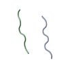
| ||||||||
|---|---|---|---|---|---|---|---|---|---|
| 1 | 
| ||||||||
| Unit cell |
| ||||||||
| Details | The biological assembly is a pair of indefinitely long beta sheets. The first sheet is composed of the following symmetry mates of Chain A: x,y,z; -x,1/2+y,1/2-z; x,y+1,z; -x,3/2+y,1/2-z; x,y+2,z; etc. The complementary sheet is composed of the following symmetry mates of Chain B: x,y,z; -x,1/2+y,-1/2-z; x,y+1,z; -x,3/2+y,-1/2-z; x,y+2,z; etc |
-Components
| #1: Protein/peptide | Mass: 664.746 Da / Num. of mol.: 2 / Source method: obtained synthetically Details: This sequence corresponds to a segment of human transthyretin Source: (synth.) synthetic construct (others) #2: Water | ChemComp-HOH / |  Mass: 18.015 Da / Num. of mol.: 6 / Source method: isolated from a natural source / Formula: H2O Mass: 18.015 Da / Num. of mol.: 6 / Source method: isolated from a natural source / Formula: H2O |
|---|
-Experimental details
-Experiment
| Experiment | Method: X-RAY DIFFRACTION |
|---|
- Sample preparation
| Crystal | Density Matthews: 1.44 Å3/Da / Density % sol: 14.6 % |
|---|---|
| Crystal grow | Temperature: 298 K / Method: vapor diffusion, hanging drop / Details: 0.4M Ammonium dihydrogen phosphate, 25% Glycerol |
-Data collection
| Diffraction | Mean temperature: 100 K |
|---|---|
| Diffraction source | Source: SYNCHROTRON / Site: APS  / Beamline: 24-ID-E / Wavelength: 0.9792 Å / Beamline: 24-ID-E / Wavelength: 0.9792 Å |
| Detector | Type: PSI PILATUS 6M / Detector: PIXEL / Date: Aug 9, 2013 |
| Radiation | Monochromator: Si (111) / Protocol: SINGLE WAVELENGTH / Monochromatic (M) / Laue (L): M / Scattering type: x-ray |
| Radiation wavelength | Wavelength: 0.9792 Å / Relative weight: 1 |
| Reflection | Resolution: 1.85→14.117 Å / Num. obs: 740 / % possible obs: 93.7 % / Redundancy: 23.1 % / Net I/σ(I): 4.71 |
- Processing
| Software |
| ||||||||||||||||||||||||
|---|---|---|---|---|---|---|---|---|---|---|---|---|---|---|---|---|---|---|---|---|---|---|---|---|---|
| Refinement | Method to determine structure: MOLECULAR REPLACEMENT / Resolution: 1.85→14.117 Å / SU ML: 0.15 / Cross valid method: FREE R-VALUE / σ(F): 1.39 / Phase error: 15.64 / Stereochemistry target values: ML
| ||||||||||||||||||||||||
| Solvent computation | Shrinkage radii: 0.9 Å / VDW probe radii: 1.11 Å / Solvent model: FLAT BULK SOLVENT MODEL | ||||||||||||||||||||||||
| Refinement step | Cycle: LAST / Resolution: 1.85→14.117 Å
| ||||||||||||||||||||||||
| Refine LS restraints |
|
