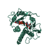
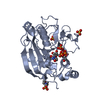
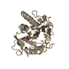
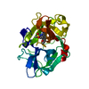
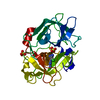
[English] 日本語
Yorodumi
- PDB-4xe4: Coagulation Factor XII protease domain crystal structure -

+
Open data


ID or keywords:

Loading...

-
Basic information

Entry
Database: PDB / ID: 4xe4
TitleCoagulation Factor XII protease domain crystal structure
ComponentsCoagulation factor XII
KeywordsBLOOD CLOTTING / Factor XII / Zymogens / Catalytic Domain / Active site
Function / homology
Function and homology information


coagulation factor XIIa / plasma kallikrein-kinin cascade / Factor XII activation / Defective SERPING1 causes hereditary angioedema / response to misfolded protein / positive regulation of plasminogen activation / blood coagulation, intrinsic pathway / positive regulation of fibrinolysis / misfolded protein binding / zymogen activation ...coagulation factor XIIa / plasma kallikrein-kinin cascade / Factor XII activation / Defective SERPING1 causes hereditary angioedema / response to misfolded protein / positive regulation of plasminogen activation / blood coagulation, intrinsic pathway / positive regulation of fibrinolysis / misfolded protein binding / zymogen activation / Defective factor XII causes hereditary angioedema / protein autoprocessing / positive regulation of blood coagulation / rough endoplasmic reticulum / fibrinolysis / Intrinsic Pathway of Fibrin Clot Formation / protein processing / blood coagulation / : / innate immune response / serine-type endopeptidase activity / calcium ion binding / extracellular space / extracellular exosome / extracellular region / plasma membrane
Similarity search - Function
Coagulation factor XII/hepatocyte growth factor activator / Fibronectin type I domain / Fibronectin, type I / Fibronectin type-I domain signature. / Fibronectin type-I domain profile. / Fibronectin type 1 domain / Fibronectin type II domain / Fibronectin type II domain superfamily / Fibronectin type II domain / Fibronectin type-II collagen-binding domain signature. ...Coagulation factor XII/hepatocyte growth factor activator / Fibronectin type I domain / Fibronectin, type I / Fibronectin type-I domain signature. / Fibronectin type-I domain profile. / Fibronectin type 1 domain / Fibronectin type II domain / Fibronectin type II domain superfamily / Fibronectin type II domain / Fibronectin type-II collagen-binding domain signature. / Fibronectin type-II collagen-binding domain profile. / Fibronectin type 2 domain / EGF-like domain / EGF-like calcium-binding domain / Calcium-binding EGF-like domain / Kringle domain / Kringle / Kringle, conserved site / Kringle superfamily / Kringle domain signature. / Kringle domain profile. / Kringle domain / : / Kringle-like fold / Epidermal growth factor-like domain. / EGF-like domain profile. / EGF-like domain signature 1. / EGF-like domain signature 2. / EGF-like domain / Serine proteases, trypsin family, histidine active site / Serine proteases, trypsin family, serine active site / Serine proteases, trypsin family, histidine active site. / Peptidase S1A, chymotrypsin family / Serine proteases, trypsin family, serine active site. / Serine proteases, trypsin domain profile. / Trypsin-like serine protease / Serine proteases, trypsin domain / Trypsin / Trypsin-like serine proteases / Thrombin, subunit H / Peptidase S1, PA clan, chymotrypsin-like fold / Peptidase S1, PA clan / Beta Barrel / Mainly Beta
Similarity search - Domain/homology
triacetyl-beta-chitotriose / Coagulation factor XII
Similarity search - Component
Biological speciesHomo sapiens (human)
MethodX-RAY DIFFRACTION / SYNCHROTRON / MOLECULAR REPLACEMENT / Resolution: 2.4 Å
AuthorsPathak, M. / Wilmann, P. / Awford, J. / Li, C. / Fisher, P.M. / Dreveny, I. / Dekker, L.V. / Emsley, J.
Funding support United Kingdom, 1items
OrganizationGrant numberCountry
United Kingdom
CitationJournal: J.Thromb.Haemost. / Year: 2015
Title: Coagulation factor XII protease domain crystal structure.
Authors: Pathak, M. / Wilmann, P. / Awford, J. / Li, C. / Hamad, B.K. / Fischer, P.M. / Dreveny, I. / Dekker, L.V. / Emsley, J.
History
DepositionDec 22, 2014Deposition site: RCSB / Processing site: PDBE
Revision 1.0Feb 4, 2015Provider: repository / Type: Initial release
Revision 1.1Apr 15, 2015Group: Database references
Revision 1.2Oct 28, 2015Group: Database references
Revision 2.0Jul 29, 2020Group: Advisory / Atomic model ...Advisory / Atomic model / Data collection / Derived calculations / Structure summary
Category: atom_site / chem_comp ...atom_site / chem_comp / entity / entity_name_com / pdbx_branch_scheme / pdbx_chem_comp_identifier / pdbx_entity_branch / pdbx_entity_branch_descriptor / pdbx_entity_branch_link / pdbx_entity_branch_list / pdbx_entity_nonpoly / pdbx_molecule_features / pdbx_nonpoly_scheme / pdbx_struct_assembly_gen / pdbx_struct_special_symmetry / pdbx_unobs_or_zero_occ_atoms / struct_asym / struct_conn / struct_site / struct_site_gen
Item: _atom_site.B_iso_or_equiv / _atom_site.Cartn_x ..._atom_site.B_iso_or_equiv / _atom_site.Cartn_x / _atom_site.Cartn_y / _atom_site.Cartn_z / _atom_site.auth_asym_id / _atom_site.auth_atom_id / _atom_site.auth_seq_id / _atom_site.label_asym_id / _atom_site.label_atom_id / _atom_site.type_symbol / _chem_comp.name / _chem_comp.type / _entity.formula_weight / _entity.pdbx_description / _entity.pdbx_number_of_molecules / _entity.type / _pdbx_struct_assembly_gen.asym_id_list / _pdbx_struct_special_symmetry.label_asym_id / _struct_conn.pdbx_dist_value / _struct_conn.pdbx_leaving_atom_flag / _struct_conn.pdbx_role / _struct_conn.pdbx_value_order / _struct_conn.ptnr1_auth_asym_id / _struct_conn.ptnr1_auth_seq_id / _struct_conn.ptnr1_label_asym_id / _struct_conn.ptnr1_label_atom_id / _struct_conn.ptnr2_auth_asym_id / _struct_conn.ptnr2_auth_seq_id / _struct_conn.ptnr2_label_asym_id / _struct_conn.ptnr2_label_atom_id
Description: Carbohydrate remediation / Provider: repository / Type: Remediation
Revision 2.1Jan 10, 2024Group: Advisory / Data collection ...Advisory / Data collection / Database references / Refinement description / Structure summary
Category: chem_comp / chem_comp_atom ...chem_comp / chem_comp_atom / chem_comp_bond / database_2 / pdbx_initial_refinement_model / pdbx_unobs_or_zero_occ_atoms
Item: _chem_comp.pdbx_synonyms / _database_2.pdbx_DOI / _database_2.pdbx_database_accession
Revision 2.2Nov 6, 2024Group: Structure summary / Category: pdbx_entry_details / pdbx_modification_feature

-
Structure visualization

Structure viewerMolecule:
MolmilJmol/JSmol

Downloads & links

-
Assembly

Deposited unit
A: Coagulation factor XII
hetero molecules


Theoretical massNumber of molelcules
Total (without water)28,3533
Polymers27,6331
Non-polymers7202
Water1,40578
1


  • Idetical with deposited unit
  • defined by author&software
  • MONOMERIC
TypeNameSymmetry operationNumber
identity operation1_555x,y,z1
Buried area1160 Å2
ΔGint13 kcal/mol
Surface area11670 Å2
MethodPISA
Unit cell
Length a, b, c (Å)137.060, 137.060, 37.011
Angle α, β, γ (deg.)90.00, 90.00, 120.00
Int Tables number154
Space group name H-MP3221
Components on special symmetry positions
IDModelComponents
11A-808-

HOH

21A-872-

HOH

-
Components

#1: Protein Coagulation factor XII / Hageman factor / HAF


Mass: 27632.955 Da / Num. of mol.: 1 / Mutation: C467S
Source method: isolated from a genetically manipulated source
Source: (gene. exp.) Homo sapiens (human) / Gene: F12 / Plasmid: pMT-PURO / Cell line (production host): Schneider 2 (S2) Cells / Production host: Drosophila melanogaster (fruit fly) / References: UniProt: P00748, coagulation factor XIIa
#2: Polysaccharide 2-acetamido-2-deoxy-beta-D-glucopyranose-(1-4)-2-acetamido-2-deoxy-beta-D-glucopyranose-(1-4)-2- ...2-acetamido-2-deoxy-beta-D-glucopyranose-(1-4)-2-acetamido-2-deoxy-beta-D-glucopyranose-(1-4)-2-acetamido-2-deoxy-beta-D-glucopyranose / triacetyl-beta-chitotriose


Type: oligosaccharide, Oligosaccharide / Class: Inhibitor / Mass: 627.594 Da / Num. of mol.: 1
Source method: isolated from a genetically manipulated source
Details: oligosaccharide / References: triacetyl-beta-chitotriose
DescriptorTypeProgram
DGlcpNAcb1-4DGlcpNAcb1-4DGlcpNAcb1-Glycam Condensed SequenceGMML 1.0
WURCS=2.0/1,3,2/[a2122h-1b_1-5_2*NCC/3=O]/1-1-1/a4-b1_b4-c1WURCSPDB2Glycan 1.1.0
[]{[(4+1)][b-D-GlcpNAc]{[(4+1)][b-D-GlcpNAc]{[(4+1)][b-D-GlcpNAc]{}}}}LINUCSPDB-CARE
#3: Chemical ChemComp-GOL / GLYCEROL / GLYCERIN / PROPANE-1,2,3-TRIOL


Mass: 92.094 Da / Num. of mol.: 1 / Source method: obtained synthetically / Formula: C3H8O3
#4: Water ChemComp-HOH / water


Mass: 18.015 Da / Num. of mol.: 78 / Source method: isolated from a natural source / Formula: H2O
Has protein modificationY

-
Experimental details

-
Experiment

ExperimentMethod: X-RAY DIFFRACTION

-
Sample preparation

CrystalDensity Matthews: 3.64 Å3/Da / Density % sol: 66.19 %
Crystal growTemperature: 293 K / Method: vapor diffusion, hanging drop
Details: 0.1M Hepes pH 7.5,1.6M Ammonium sulphate, 2%(w/v) PEG1000.

-
Data collection

DiffractionMean temperature: 100 K
Diffraction sourceSource: SYNCHROTRON / Site: Diamond / Beamline: I02 / Wavelength: 0.97949 Å
DetectorType: ADSC QUANTUM 315r / Detector: CCD / Date: Oct 18, 2014
RadiationProtocol: SINGLE WAVELENGTH / Monochromatic (M) / Laue (L): M / Scattering type: x-ray
Radiation wavelengthWavelength: 0.97949 Å / Relative weight: 1
ReflectionResolution: 2.39→39.57 Å / Num. all: 15737 / Num. obs: 15737 / % possible obs: 98.4 % / Redundancy: 5.6 % / Biso Wilson estimate: 59.9 Å2 / Rmerge(I) obs: 0.069 / Net I/σ(I): 11
Reflection shellResolution: 2.39→2.48 Å / Redundancy: 5.7 % / Mean I/σ(I) obs: 2.2 / % possible all: 99

-
Processing

Software
NameVersionClassification
REFMAC5.7.0029refinement
MOSFLMdata reduction
Aimlessdata scaling
PHASERphasing
RefinementMethod to determine structure: MOLECULAR REPLACEMENT
Starting model: 4XDE
Resolution: 2.4→39.57 Å / Cor.coef. Fo:Fc: 0.953 / Cor.coef. Fo:Fc free: 0.919 / SU B: 10.772 / SU ML: 0.237 / Cross valid method: THROUGHOUT / ESU R: 0.275 / ESU R Free: 0.259 / Stereochemistry target values: MAXIMUM LIKELIHOOD / Details: HYDROGENS HAVE BEEN ADDED IN THE RIDING POSITIONS
RfactorNum. reflection% reflectionSelection details
Rfree0.29504 723 4.7 %RANDOM
Rwork0.21845 ---
obs0.2218 14815 98.16 %-
Solvent computationIon probe radii: 0.8 Å / Shrinkage radii: 0.8 Å / VDW probe radii: 1.2 Å / Solvent model: MASK
Displacement parameters
Baniso -1Baniso -2Baniso -3
1-1.28 Å21.28 Å20 Å2
2--1.28 Å20 Å2
3----4.15 Å2
Refinement stepCycle: 1 / Resolution: 2.4→39.57 Å
ProteinNucleic acidLigandSolventTotal
Num. atoms1765 0 48 78 1891
Refine LS restraints
Refine-IDTypeDev idealDev ideal targetNumber
X-RAY DIFFRACTIONr_bond_refined_d0.0160.0191866
X-RAY DIFFRACTIONr_bond_other_d0.0030.021699
X-RAY DIFFRACTIONr_angle_refined_deg1.9631.9772545
X-RAY DIFFRACTIONr_angle_other_deg0.9643.0053876
X-RAY DIFFRACTIONr_dihedral_angle_1_deg8.2495231
X-RAY DIFFRACTIONr_dihedral_angle_2_deg38.91723.37580
X-RAY DIFFRACTIONr_dihedral_angle_3_deg22.34915259
X-RAY DIFFRACTIONr_dihedral_angle_4_deg21.4861512
X-RAY DIFFRACTIONr_chiral_restr0.1030.2282
X-RAY DIFFRACTIONr_gen_planes_refined0.0090.0212111
X-RAY DIFFRACTIONr_gen_planes_other0.0010.02431
X-RAY DIFFRACTIONr_nbd_refined
X-RAY DIFFRACTIONr_nbd_other
X-RAY DIFFRACTIONr_nbtor_refined
X-RAY DIFFRACTIONr_nbtor_other
X-RAY DIFFRACTIONr_xyhbond_nbd_refined
X-RAY DIFFRACTIONr_xyhbond_nbd_other
X-RAY DIFFRACTIONr_metal_ion_refined
X-RAY DIFFRACTIONr_metal_ion_other
X-RAY DIFFRACTIONr_symmetry_vdw_refined
X-RAY DIFFRACTIONr_symmetry_vdw_other
X-RAY DIFFRACTIONr_symmetry_hbond_refined
X-RAY DIFFRACTIONr_symmetry_hbond_other
X-RAY DIFFRACTIONr_symmetry_metal_ion_refined
X-RAY DIFFRACTIONr_symmetry_metal_ion_other
X-RAY DIFFRACTIONr_mcbond_it
X-RAY DIFFRACTIONr_mcbond_other
X-RAY DIFFRACTIONr_mcangle_it
X-RAY DIFFRACTIONr_mcangle_other
X-RAY DIFFRACTIONr_scbond_it
X-RAY DIFFRACTIONr_scbond_other
X-RAY DIFFRACTIONr_scangle_it
X-RAY DIFFRACTIONr_scangle_other
X-RAY DIFFRACTIONr_long_range_B_refined
X-RAY DIFFRACTIONr_long_range_B_other
X-RAY DIFFRACTIONr_rigid_bond_restr
X-RAY DIFFRACTIONr_sphericity_free
X-RAY DIFFRACTIONr_sphericity_bonded
LS refinement shellResolution: 2.4→2.462 Å
RfactorNum. reflection% reflection
Rfree0.417 52 -
Rwork-1110 -
obs--98.64 %

+
About Yorodumi

-
News

-
Feb 9, 2022. New format data for meta-information of EMDB entries

New format data for meta-information of EMDB entries

  • Version 3 of the EMDB header file is now the official format.
  • The previous official version 1.9 will be removed from the archive.

Related info.:EMDB header

External links:wwPDB to switch to version 3 of the EMDB data model

-
Aug 12, 2020. Covid-19 info

Covid-19 info

URL: https://pdbj.org/emnavi/covid19.php

New page: Covid-19 featured information page in EM Navigator.

Related info.:Covid-19 info / Mar 5, 2020. Novel coronavirus structure data

+
Mar 5, 2020. Novel coronavirus structure data

Novel coronavirus structure data

Related info.:Yorodumi Speices / Aug 12, 2020. Covid-19 info

External links:COVID-19 featured content - PDBj / Molecule of the Month (242):Coronavirus Proteases

+
Jan 31, 2019. EMDB accession codes are about to change! (news from PDBe EMDB page)

EMDB accession codes are about to change! (news from PDBe EMDB page)

  • The allocation of 4 digits for EMDB accession codes will soon come to an end. Whilst these codes will remain in use, new EMDB accession codes will include an additional digit and will expand incrementally as the available range of codes is exhausted. The current 4-digit format prefixed with “EMD-” (i.e. EMD-XXXX) will advance to a 5-digit format (i.e. EMD-XXXXX), and so on. It is currently estimated that the 4-digit codes will be depleted around Spring 2019, at which point the 5-digit format will come into force.
  • The EM Navigator/Yorodumi systems omit the EMD- prefix.

Related info.:Q: What is EMD? / ID/Accession-code notation in Yorodumi/EM Navigator

External links:EMDB Accession Codes are Changing Soon! / Contact to PDBj

+
Jul 12, 2017. Major update of PDB

Major update of PDB

  • wwPDB released updated PDB data conforming to the new PDBx/mmCIF dictionary.
  • This is a major update changing the version number from 4 to 5, and with Remediation, in which all the entries are updated.
  • In this update, many items about electron microscopy experimental information are reorganized (e.g. em_software).
  • Now, EM Navigator and Yorodumi are based on the updated data.

External links:wwPDB Remediation / Enriched Model Files Conforming to OneDep Data Standards Now Available in the PDB FTP Archive

-
Yorodumi

Thousand views of thousand structures

  • Yorodumi is a browser for structure data from EMDB, PDB, SASBDB, etc.
  • This page is also the successor to EM Navigator detail page, and also detail information page/front-end page for Omokage search.
  • The word "yorodu" (or yorozu) is an old Japanese word meaning "ten thousand". "mi" (miru) is to see.

Related info.:EMDB / PDB / SASBDB / Comparison of 3 databanks / Yorodumi Search / Aug 31, 2016. New EM Navigator & Yorodumi / Yorodumi Papers / Jmol/JSmol / Function and homology information / Changes in new EM Navigator and Yorodumi

Read more