 Movie
Movie Controller

[English] 日本語
 Yorodumi
- PDB-4xdy: Structure of NADH-preferring ketol-acid reductoisomerase from an ... -
+ Open data

- Basic information
| Entry | Database: PDB / ID: 4xdy | ||||||
|---|---|---|---|---|---|---|---|
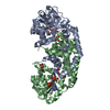
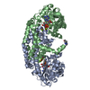
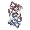
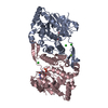
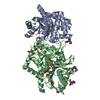
| Title | Structure of NADH-preferring ketol-acid reductoisomerase from an uncultured archean | ||||||
 Components Components | Ketol-acid reductoisomerase | ||||||
 Keywords Keywords | OXIDOREDUCTASE / Rossmann fold | ||||||
| Function / homology |  Function and homology information Function and homology informationketol-acid reductoisomerase (NAD+) / ketol-acid reductoisomerase activity / L-valine biosynthetic process / : / NADP binding / magnesium ion binding Similarity search - Function | ||||||
| Biological species |  uncultured archaeon GZfos26G2 (environmental samples) uncultured archaeon GZfos26G2 (environmental samples) | ||||||
| Method |  X-RAY DIFFRACTION / SYNCHROTRON / MOLECULAR REPLACEMENT / Resolution: 1.535 Å X-RAY DIFFRACTION / SYNCHROTRON / MOLECULAR REPLACEMENT / Resolution: 1.535 Å | ||||||
 Authors Authors | Cahn, J.K.B. / Brinkmann-Chen, S. / Arnold, F.H. | ||||||
 Citation Citation | Journal: Biochem.J. / Year: 2015 Title: Cofactor specificity motifs and the induced fit mechanism in class I ketol-acid reductoisomerases. Authors: Cahn, J.K. / Brinkmann-Chen, S. / Spatzal, T. / Wiig, J.A. / Buller, A.R. / Einsle, O. / Hu, Y. / Ribbe, M.W. / Arnold, F.H. #1: Journal: Metab. Eng. / Year: 2014Title: Uncovering rare NADH-preferring ketol-acid reductoisomerases. Authors: Brinkmann-Chen, S. / Cahn, J.K. / Arnold, F.H. | ||||||
| History |
|
- Structure visualization
| Structure viewer | Molecule: MolmilJmol/JSmol |
|---|
- Downloads & links
-Download
| PDBx/mmCIF format | 4xdy.cif.gz | 158.4 KB | Display | PDBx/mmCIF format |
|---|---|---|---|---|
| PDB format | pdb4xdy.ent.gz | 122.9 KB | Display | PDB format |
| PDBx/mmJSON format | 4xdy.json.gz | Tree view | PDBx/mmJSON format | |
| Others |  Other downloads Other downloads |
-Validation report
| Arichive directory | https://data.pdbj.org/pub/pdb/validation_reports/xd/4xdyftp://data.pdbj.org/pub/pdb/validation_reports/xd/4xdy | HTTPS FTP |
|---|
-Related structure data
| Related structure data | 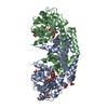 4xdzC 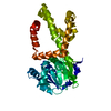 4xehC 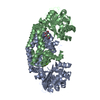 4xiyC  4kqxS S: Starting model for refinement C: citing same article ( |
|---|---|
| Similar structure data |


-Links
 PDBj
PDBj


- Assembly
| Deposited unit | 
| ||||||||
|---|---|---|---|---|---|---|---|---|---|
| 1 |
| ||||||||
| Unit cell |
|
-Components
-Protein , 1 types, 2 molecules AB
| #1: Protein | Mass: 37730.156 Da / Num. of mol.: 2 Source method: isolated from a genetically manipulated source Source: (gene. exp.) uncultured archaeon GZfos26G2 (environmental samples)Gene: ilvC, GZ26G2_30 / Plasmid: pET22b(+) / Production host:  References: UniProt: Q64BR7, ketol-acid reductoisomerase (NADP+) |
|---|
-Non-polymers , 5 types, 371 molecules 


| #2: Chemical | ChemComp-MG /  Mass: 24.305 Da / Num. of mol.: 6 Mass: 24.305 Da / Num. of mol.: 6Source method: isolated from a genetically manipulated source Formula: Mg #3: Chemical |  Mass: 665.441 Da / Num. of mol.: 2 / Source method: obtained synthetically / Formula: C21H29N7O14P2 Mass: 665.441 Da / Num. of mol.: 2 / Source method: obtained synthetically / Formula: C21H29N7O14P2#4: Chemical |  Mass: 92.094 Da / Num. of mol.: 2 / Source method: obtained synthetically / Formula: C3H8O3 Mass: 92.094 Da / Num. of mol.: 2 / Source method: obtained synthetically / Formula: C3H8O3#5: Chemical |  Mass: 147.129 Da / Num. of mol.: 2 / Source method: obtained synthetically / Formula: C5H9NO4 Mass: 147.129 Da / Num. of mol.: 2 / Source method: obtained synthetically / Formula: C5H9NO4#6: Water | ChemComp-HOH / | Mass: 18.015 Da / Num. of mol.: 359 / Source method: isolated from a natural source / Formula: H2O |
|---|
-Experimental details
-Experiment
| Experiment | Method: X-RAY DIFFRACTION |
|---|
- Sample preparation
| Crystal | Density Matthews: 3.51 Å3/Da / Density % sol: 64.98 % |
|---|---|
| Crystal grow | Temperature: 295 K / Method: vapor diffusion, sitting drop / pH: 5 Details: 0.1 M bis-tris pH 5, 20% w/v polyethylene glycol 1500 |
-Data collection
| Diffraction | Mean temperature: 100 K |
|---|---|
| Diffraction source | Source: SYNCHROTRON / Site: SSRL  / Beamline: BL12-2 / Wavelength: 0.9795 Å / Beamline: BL12-2 / Wavelength: 0.9795 Å |
| Detector | Type: DECTRIS PILATUS 6M / Detector: PIXEL / Date: Mar 30, 2013 |
| Radiation | Monochromator: LIQUID NITROGEN-COOLED DOUBLE CRYSTAL K-B FOCUSING MIRRORS Protocol: SINGLE WAVELENGTH / Monochromatic (M) / Laue (L): M / Scattering type: x-ray |
| Radiation wavelength | Wavelength: 0.9795 Å / Relative weight: 1 |
| Reflection | Resolution: 1.535→102.57 Å / Num. obs: 157419 / % possible obs: 99.4 % / Redundancy: 4.5 % / Rsym value: 0.088 / Net I/σ(I): 7.2 |
| Reflection shell | Resolution: 1.535→1.7 Å / Redundancy: 4.5 % / Rmerge(I) obs: 0.963 / Mean I/σ(I) obs: 1.5 / % possible all: 99.8 |
- Processing
| Software |
| ||||||||||||||||||||||||||||||||||||||||||||||||||||||||||||||||||||||||||||||||||||||||||||||||||||||||||||||||||||||||||||||||||||||||||||||||||||||||||||||||||||||||||||||||||||||
|---|---|---|---|---|---|---|---|---|---|---|---|---|---|---|---|---|---|---|---|---|---|---|---|---|---|---|---|---|---|---|---|---|---|---|---|---|---|---|---|---|---|---|---|---|---|---|---|---|---|---|---|---|---|---|---|---|---|---|---|---|---|---|---|---|---|---|---|---|---|---|---|---|---|---|---|---|---|---|---|---|---|---|---|---|---|---|---|---|---|---|---|---|---|---|---|---|---|---|---|---|---|---|---|---|---|---|---|---|---|---|---|---|---|---|---|---|---|---|---|---|---|---|---|---|---|---|---|---|---|---|---|---|---|---|---|---|---|---|---|---|---|---|---|---|---|---|---|---|---|---|---|---|---|---|---|---|---|---|---|---|---|---|---|---|---|---|---|---|---|---|---|---|---|---|---|---|---|---|---|---|---|---|---|
| Refinement | Method to determine structure: MOLECULAR REPLACEMENT Starting model: 4KQX Resolution: 1.535→102.57 Å / Cor.coef. Fo:Fc: 0.972 / Cor.coef. Fo:Fc free: 0.959 / SU B: 1.989 / SU ML: 0.064 / Cross valid method: THROUGHOUT / ESU R: 0.061 / ESU R Free: 0.066 / Stereochemistry target values: MAXIMUM LIKELIHOOD / Details: HYDROGENS HAVE BEEN ADDED IN THE RIDING POSITIONS
| ||||||||||||||||||||||||||||||||||||||||||||||||||||||||||||||||||||||||||||||||||||||||||||||||||||||||||||||||||||||||||||||||||||||||||||||||||||||||||||||||||||||||||||||||||||||
| Solvent computation | Ion probe radii: 0.8 Å / Shrinkage radii: 0.8 Å / VDW probe radii: 1.2 Å / Solvent model: MASK | ||||||||||||||||||||||||||||||||||||||||||||||||||||||||||||||||||||||||||||||||||||||||||||||||||||||||||||||||||||||||||||||||||||||||||||||||||||||||||||||||||||||||||||||||||||||
| Displacement parameters | Biso mean: 25.89 Å2
| ||||||||||||||||||||||||||||||||||||||||||||||||||||||||||||||||||||||||||||||||||||||||||||||||||||||||||||||||||||||||||||||||||||||||||||||||||||||||||||||||||||||||||||||||||||||
| Refinement step | Cycle: LAST / Resolution: 1.535→102.57 Å
| ||||||||||||||||||||||||||||||||||||||||||||||||||||||||||||||||||||||||||||||||||||||||||||||||||||||||||||||||||||||||||||||||||||||||||||||||||||||||||||||||||||||||||||||||||||||
| Refine LS restraints |
|