 Movie
Movie Controller

[English] 日本語
 Yorodumi
- PDB-4x0t: Structure ALDH7A1 inactivated by 4-diethylaminobenzaldehyde and c... -
+ Open data

- Basic information
| Entry | Database: PDB / ID: 4x0t | ||||||
|---|---|---|---|---|---|---|---|
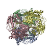
| Title | Structure ALDH7A1 inactivated by 4-diethylaminobenzaldehyde and complexed with NAD+ | ||||||
 Components Components | Alpha-aminoadipic semialdehyde dehydrogenase | ||||||
 Keywords Keywords | oxidoreductase/oxidoreductase inhibitor / ALDEHYDE DEHYDROGENASE / OXIDOREDUCTASE / LYSINE CATABOLISM / oxidoreductase-oxidoreductase inhibitor complex | ||||||
| Function / homology |  Function and homology information Function and homology informationnegative regulation of endosome to plasma membrane protein transport / L-aminoadipate-semialdehyde dehydrogenase [NAD(P)+] activity / L-aminoadipate-semialdehyde dehydrogenase / Choline catabolism / negative regulation of Golgi to plasma membrane protein transport / L-lysine catabolic process / choline catabolic process / Lysine catabolism / : / betaine-aldehyde dehydrogenase (NAD+) activity ...negative regulation of endosome to plasma membrane protein transport / L-aminoadipate-semialdehyde dehydrogenase [NAD(P)+] activity / L-aminoadipate-semialdehyde dehydrogenase / Choline catabolism / negative regulation of Golgi to plasma membrane protein transport / L-lysine catabolic process / choline catabolic process / Lysine catabolism / : / betaine-aldehyde dehydrogenase (NAD+) activity / betaine-aldehyde dehydrogenase / : / aldehyde metabolic process / plasma membrane to endosome transport / aldehyde dehydrogenase (NAD+) / Golgi to endosome transport / aldehyde dehydrogenase (NAD+) activity / oxidoreductase activity, acting on NAD(P)H, quinone or similar compound as acceptor / endosome to lysosome transport / negative regulation of ferroptosis / energy homeostasis / sensory perception of sound / mitochondrial matrix / Golgi membrane / mitochondrion / extracellular exosome / identical protein binding / nucleus / plasma membrane / cytosol Similarity search - Function | ||||||
| Biological species |  Homo sapiens (human) Homo sapiens (human) | ||||||
| Method |  X-RAY DIFFRACTION / SYNCHROTRON / MOLECULAR REPLACEMENT / Resolution: 2.4 Å X-RAY DIFFRACTION / SYNCHROTRON / MOLECULAR REPLACEMENT / Resolution: 2.4 Å | ||||||
 Authors Authors | Luo, M. / Tanner, J.J. | ||||||
 Citation Citation | Journal: Acs Chem.Biol. / Year: 2015 Title: Diethylaminobenzaldehyde Is a Covalent, Irreversible Inactivator of ALDH7A1. Authors: Luo, M. / Gates, K.S. / Henzl, M.T. / Tanner, J.J. | ||||||
| History |
|
- Structure visualization
| Structure viewer | Molecule: MolmilJmol/JSmol |
|---|
- Downloads & links
-Download
| PDBx/mmCIF format | 4x0t.cif.gz | 758.3 KB | Display | PDBx/mmCIF format |
|---|---|---|---|---|
| PDB format | pdb4x0t.ent.gz | 633.7 KB | Display | PDB format |
| PDBx/mmJSON format | 4x0t.json.gz | Tree view | PDBx/mmJSON format | |
| Others |  Other downloads Other downloads |
-Validation report
| Arichive directory | https://data.pdbj.org/pub/pdb/validation_reports/x0/4x0tftp://data.pdbj.org/pub/pdb/validation_reports/x0/4x0t | HTTPS FTP |
|---|
-Related structure data
| Related structure data |  4x0uC  2j6lS C: citing same article ( S: Starting model for refinement |
|---|---|
| Similar structure data |


-Links
 PDBj
PDBj

- Assembly
| Deposited unit | 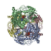
| ||||||||
|---|---|---|---|---|---|---|---|---|---|
| 1 |
| ||||||||
| Unit cell |
| ||||||||
| Components on special symmetry positions |
| ||||||||
| Details | tetramer |
-Components
| #1: Protein | Mass: 55620.367 Da / Num. of mol.: 4 Source method: isolated from a genetically manipulated source Source: (gene. exp.) Homo sapiens (human) / Gene: ALDH7A1, ATQ1 / Production host:  References: UniProt: P49419, L-aminoadipate-semialdehyde dehydrogenase, aldehyde dehydrogenase (NAD+), betaine-aldehyde dehydrogenase #2: Chemical | ChemComp-NAD /   Mass: 663.425 Da / Num. of mol.: 4 / Source method: obtained synthetically / Formula: C21H27N7O14P2 / Comment: NAD*YM Mass: 663.425 Da / Num. of mol.: 4 / Source method: obtained synthetically / Formula: C21H27N7O14P2 / Comment: NAD*YM#3: Chemical | ChemComp-3W9 /   Mass: 177.243 Da / Num. of mol.: 4 / Source method: obtained synthetically / Formula: C11H15NO Mass: 177.243 Da / Num. of mol.: 4 / Source method: obtained synthetically / Formula: C11H15NO#4: Water | ChemComp-HOH / |  Mass: 18.015 Da / Num. of mol.: 314 / Source method: isolated from a natural source / Formula: H2O Mass: 18.015 Da / Num. of mol.: 314 / Source method: isolated from a natural source / Formula: H2OHas protein modification | Y | |
|---|
-Experimental details
-Experiment
| Experiment | Method: X-RAY DIFFRACTION / Number of used crystals: 1 |
|---|
- Sample preparation
| Crystal | Density Matthews: 2.31 Å3/Da / Density % sol: 46.81 % |
|---|---|
| Crystal grow | Temperature: 293 K / Method: vapor diffusion, sitting drop Details: The reservoir contained 0.1 M magnesium formate dihydrate, 15% w/v PEG 3350 and 0.1 M Hepes. The enzyme stock solution included 3 mg/mL ALDH7A1, 200 micromolar 4-diethyaminobenzaldehyde and 5 mM NAD+. PH range: 7.5 |
-Data collection
| Diffraction | Mean temperature: 100 K |
|---|---|
| Diffraction source | Source: SYNCHROTRON / Site: ALS  / Beamline: 4.2.2 / Wavelength: 1 Å / Beamline: 4.2.2 / Wavelength: 1 Å |
| Detector | Type: CUSTOM-MADE / Detector: CMOS / Date: Aug 22, 2014 / Details: Taurus-1 detector |
| Radiation | Protocol: SINGLE WAVELENGTH / Monochromatic (M) / Laue (L): M / Scattering type: x-ray |
| Radiation wavelength | Wavelength: 1 Å / Relative weight: 1 |
| Reflection | Resolution: 2.4→59.52 Å / Num. obs: 81067 / % possible obs: 99.7 % / Redundancy: 7.5 % / Biso Wilson estimate: 30.27 Å2 / Rmerge(I) obs: 0.154 / Net I/σ(I): 12 |
| Reflection shell | Resolution: 2.4→2.45 Å / Redundancy: 7.5 % / Rmerge(I) obs: 1.063 / Mean I/σ(I) obs: 1.9 / % possible all: 95.4 |
- Processing
| Software |
| ||||||||||||||||||||||||||||||||||||||||||||||||||||||||||||||||||||||||||||||||||||||||||||||||||||||||||||||||||||||||||||||||||||||||||||||||||||||||||||||||||||||||||||||||||||||||||||||||||||||||||||||||||
|---|---|---|---|---|---|---|---|---|---|---|---|---|---|---|---|---|---|---|---|---|---|---|---|---|---|---|---|---|---|---|---|---|---|---|---|---|---|---|---|---|---|---|---|---|---|---|---|---|---|---|---|---|---|---|---|---|---|---|---|---|---|---|---|---|---|---|---|---|---|---|---|---|---|---|---|---|---|---|---|---|---|---|---|---|---|---|---|---|---|---|---|---|---|---|---|---|---|---|---|---|---|---|---|---|---|---|---|---|---|---|---|---|---|---|---|---|---|---|---|---|---|---|---|---|---|---|---|---|---|---|---|---|---|---|---|---|---|---|---|---|---|---|---|---|---|---|---|---|---|---|---|---|---|---|---|---|---|---|---|---|---|---|---|---|---|---|---|---|---|---|---|---|---|---|---|---|---|---|---|---|---|---|---|---|---|---|---|---|---|---|---|---|---|---|---|---|---|---|---|---|---|---|---|---|---|---|---|---|---|---|---|
| Refinement | Method to determine structure: MOLECULAR REPLACEMENT Starting model: 2J6L Resolution: 2.4→59.52 Å / SU ML: 0.29 / Cross valid method: THROUGHOUT / σ(F): 1.36 / Phase error: 24.06 / Stereochemistry target values: ML
| ||||||||||||||||||||||||||||||||||||||||||||||||||||||||||||||||||||||||||||||||||||||||||||||||||||||||||||||||||||||||||||||||||||||||||||||||||||||||||||||||||||||||||||||||||||||||||||||||||||||||||||||||||
| Solvent computation | Shrinkage radii: 0.9 Å / VDW probe radii: 1.11 Å / Solvent model: FLAT BULK SOLVENT MODEL | ||||||||||||||||||||||||||||||||||||||||||||||||||||||||||||||||||||||||||||||||||||||||||||||||||||||||||||||||||||||||||||||||||||||||||||||||||||||||||||||||||||||||||||||||||||||||||||||||||||||||||||||||||
| Displacement parameters | Biso mean: 38 Å2 | ||||||||||||||||||||||||||||||||||||||||||||||||||||||||||||||||||||||||||||||||||||||||||||||||||||||||||||||||||||||||||||||||||||||||||||||||||||||||||||||||||||||||||||||||||||||||||||||||||||||||||||||||||
| Refinement step | Cycle: LAST / Resolution: 2.4→59.52 Å
| ||||||||||||||||||||||||||||||||||||||||||||||||||||||||||||||||||||||||||||||||||||||||||||||||||||||||||||||||||||||||||||||||||||||||||||||||||||||||||||||||||||||||||||||||||||||||||||||||||||||||||||||||||
| Refine LS restraints |
| ||||||||||||||||||||||||||||||||||||||||||||||||||||||||||||||||||||||||||||||||||||||||||||||||||||||||||||||||||||||||||||||||||||||||||||||||||||||||||||||||||||||||||||||||||||||||||||||||||||||||||||||||||
| LS refinement shell |
| ||||||||||||||||||||||||||||||||||||||||||||||||||||||||||||||||||||||||||||||||||||||||||||||||||||||||||||||||||||||||||||||||||||||||||||||||||||||||||||||||||||||||||||||||||||||||||||||||||||||||||||||||||
| Refinement TLS params. | Method: refined / Refine-ID: X-RAY DIFFRACTION
| ||||||||||||||||||||||||||||||||||||||||||||||||||||||||||||||||||||||||||||||||||||||||||||||||||||||||||||||||||||||||||||||||||||||||||||||||||||||||||||||||||||||||||||||||||||||||||||||||||||||||||||||||||
| Refinement TLS group |
|
