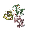
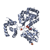
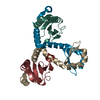
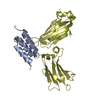
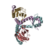
登録情報 データベース : PDB / ID : 4wwiタイトル Crystal structure of the C domain of staphylococcal protein A in complex with the Fc fragment of human IgG at 2.3 Angstrom resolution Ig gamma-3 chain C region Immunoglobulin G-binding protein A キーワード / / / / 機能・相同性 分子機能 ドメイン・相同性 構成要素
/ / / / / / / / / / / / / / / / / / / / / / / / / / / / / / / / / / / / / / / / / / / / / / / / / / / / / / / / / / / / / / / / / / / / / / / / 生物種 Staphylococcus aureus (黄色ブドウ球菌)Homo sapiens (ヒト)手法 / / / 解像度 : 2.307 Å データ登録者 Deis, L.N. / Oas, T.G. 資金援助 組織 認可番号 国 National Institutes of Health/National Institute of General Medical Sciences (NIH/NIGMS) R01-GM081666
ジャーナル : Proc.Natl.Acad.Sci.USA / 年 : 2015タイトル : Suppression of conformational heterogeneity at a protein-protein interface.著者 : Deis, L.N. / Wu, Q. / Wang, Y. / Qi, Y. / Daniels, K.G. / Zhou, P. / Oas, T.G. 履歴 登録 2014年11月11日 登録サイト / 処理サイト 改定 1.0 2015年7月15日 Provider / タイプ 改定 1.1 2015年7月22日 Group 改定 1.2 2015年7月29日 Group 改定 1.3 2017年9月13日 Group Advisory / Author supporting evidence ... Advisory / Author supporting evidence / Database references / Derived calculations / Source and taxonomy カテゴリ citation / entity_src_gen ... citation / entity_src_gen / pdbx_audit_support / pdbx_struct_oper_list / pdbx_validate_close_contact Item _citation.journal_id_CSD / _entity_src_gen.pdbx_alt_source_flag ... _citation.journal_id_CSD / _entity_src_gen.pdbx_alt_source_flag / _pdbx_audit_support.funding_organization / _pdbx_struct_oper_list.symmetry_operation 改定 1.4 2019年12月25日 Group / カテゴリ / Item 改定 1.5 2023年9月27日 Group / Database references / Refinement descriptionカテゴリ chem_comp_atom / chem_comp_bond ... chem_comp_atom / chem_comp_bond / database_2 / pdbx_initial_refinement_model Item / _database_2.pdbx_database_accession改定 1.6 2024年10月16日 Group カテゴリ / pdbx_modification_feature
すべて表示 表示を減らす
 ムービー
ムービー コントローラー


 データを開く
 基本情報
基本情報 要素
要素 キーワード
キーワード 機能・相同性情報
 Staphylococcus aureus (黄色ブドウ球菌)
 Homo sapiens (ヒト)
 X線回折 /
  データ登録者
 米国, 1件
  引用
引用 構造の表示
構造の表示 ダウンロードとリンク
 その他のダウンロード


 PDBj
PDBj


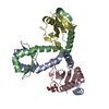
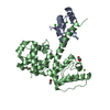
 集合体
集合体


 分子量: 18.015 Da / 分子数: 165 / 由来タイプ: 天然 / 式: H2O
 試料調製
試料調製 解析
解析