 Movie
Movie Controller

[English] 日本語
 Yorodumi
- PDB-4wwi: Crystal structure of the C domain of staphylococcal protein A in ... -
+ Open data

- Basic information
| Entry | Database: PDB / ID: 4wwi | ||||||
|---|---|---|---|---|---|---|---|
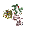
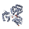
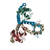
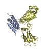
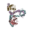
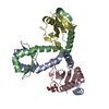
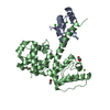
| Title | Crystal structure of the C domain of staphylococcal protein A in complex with the Fc fragment of human IgG at 2.3 Angstrom resolution | ||||||
 Components Components |
| ||||||
 Keywords Keywords | PROTEIN BINDING / three-helix bundle / conformational heterogeneity / S. aureus / antibody binding | ||||||
| Function / homology |  Function and homology information Function and homology informationIgG immunoglobulin complex / IgG binding / immunoglobulin receptor binding / immunoglobulin complex, circulating / Classical antibody-mediated complement activation / Initial triggering of complement / FCGR activation / complement activation, classical pathway / Role of phospholipids in phagocytosis / antigen binding ...IgG immunoglobulin complex / IgG binding / immunoglobulin receptor binding / immunoglobulin complex, circulating / Classical antibody-mediated complement activation / Initial triggering of complement / FCGR activation / complement activation, classical pathway / Role of phospholipids in phagocytosis / antigen binding / FCGR3A-mediated IL10 synthesis / Regulation of Complement cascade / B cell receptor signaling pathway / FCGR3A-mediated phagocytosis / Regulation of actin dynamics for phagocytic cup formation / antibacterial humoral response / blood microparticle / adaptive immune response / extracellular space / extracellular exosome / extracellular region / plasma membrane Similarity search - Function | ||||||
| Biological species |   Staphylococcus aureus (bacteria) Staphylococcus aureus (bacteria) Homo sapiens (human) Homo sapiens (human) | ||||||
| Method |  X-RAY DIFFRACTION / SYNCHROTRON / MOLECULAR REPLACEMENT / Resolution: 2.307 Å X-RAY DIFFRACTION / SYNCHROTRON / MOLECULAR REPLACEMENT / Resolution: 2.307 Å | ||||||
 Authors Authors | Deis, L.N. / Oas, T.G. | ||||||
| Funding support |  United States, 1items United States, 1items
| ||||||
 Citation Citation | Journal: Proc.Natl.Acad.Sci.USA / Year: 2015 Title: Suppression of conformational heterogeneity at a protein-protein interface. Authors: Deis, L.N. / Wu, Q. / Wang, Y. / Qi, Y. / Daniels, K.G. / Zhou, P. / Oas, T.G. | ||||||
| History |
|
- Structure visualization
| Structure viewer | Molecule: MolmilJmol/JSmol |
|---|
- Downloads & links
-Download
| PDBx/mmCIF format | 4wwi.cif.gz | 299.7 KB | Display | PDBx/mmCIF format |
|---|---|---|---|---|
| PDB format | pdb4wwi.ent.gz | 248.4 KB | Display | PDB format |
| PDBx/mmJSON format | 4wwi.json.gz | Tree view | PDBx/mmJSON format | |
| Others |  Other downloads Other downloads |
-Validation report
| Summary document | 4wwi_validation.pdf.gz | 475.5 KB | Display | wwPDB validaton report |
|---|---|---|---|---|
| Full document | 4wwi_full_validation.pdf.gz | 482 KB | Display | |
| Data in XML | 4wwi_validation.xml.gz | 28.2 KB | Display | |
| Data in CIF | 4wwi_validation.cif.gz | 39.4 KB | Display | |
| Arichive directory | https://data.pdbj.org/pub/pdb/validation_reports/ww/4wwiftp://data.pdbj.org/pub/pdb/validation_reports/ww/4wwi | HTTPS FTP |
-Related structure data
| Related structure data |  4zmdC  4zncC  1fc1S  4npdS C: citing same article ( S: Starting model for refinement |
|---|---|
| Similar structure data |


-Links
 PDBj
PDBj


- Assembly
| Deposited unit | 
| ||||||||
|---|---|---|---|---|---|---|---|---|---|
| 1 | 
| ||||||||
| 2 | 
| ||||||||
| 3 | 
| ||||||||
| Unit cell |
|
-Components
| #1: Antibody | Mass: 6637.353 Da / Num. of mol.: 3 / Fragment: UNP residues 270-327 Source method: isolated from a genetically manipulated source Source: (gene. exp.) Staphylococcus aureus (bacteria) / Gene: spa / Plasmid: pAED4 / Production host: #2: Protein | Mass: 25124.361 Da / Num. of mol.: 3 / Fragment: UNP residues 168-377 Source method: isolated from a genetically manipulated source Source: (gene. exp.) Homo sapiens (human) / Gene: IGHG3 / Production host: #3: Water | ChemComp-HOH / |  Mass: 18.015 Da / Num. of mol.: 165 / Source method: isolated from a natural source / Formula: H2O Mass: 18.015 Da / Num. of mol.: 165 / Source method: isolated from a natural source / Formula: H2OHas protein modification | Y | |
|---|
-Experimental details
-Experiment
| Experiment | Method: X-RAY DIFFRACTION / Number of used crystals: 1 |
|---|
- Sample preparation
| Crystal | Density Matthews: 3.24 Å3/Da / Density % sol: 62.09 % |
|---|---|
| Crystal grow | Temperature: 298 K / Method: vapor diffusion, sitting drop / pH: 8 / Details: NaOAc, ammonium sulfate, PEG 5K MME, glycerol |
-Data collection
| Diffraction | Mean temperature: 100 K |
|---|---|
| Diffraction source | Source: SYNCHROTRON / Site: APS / Beamline: 22-ID / Wavelength: 1 Å |
| Detector | Type: MAR CCD 165 mm / Detector: CCD / Date: Jul 19, 2014 |
| Radiation | Protocol: SINGLE WAVELENGTH / Monochromatic (M) / Laue (L): M / Scattering type: x-ray |
| Radiation wavelength | Wavelength: 1 Å / Relative weight: 1 |
| Reflection | Resolution: 2.307→41.18 Å / Num. obs: 53496 / % possible obs: 99.47 % / Redundancy: 7.5 % / Net I/σ(I): 19.5 |
| Reflection shell | Resolution: 2.307→2.389 Å / Redundancy: 6.3 % / Mean I/σ(I) obs: 1.5 / % possible all: 94.81 |
- Processing
| Software |
| ||||||||||||||||||||||||||||||||||||||||||||||||||||||||||||||||||||||||||||||||||||||||||||||||||
|---|---|---|---|---|---|---|---|---|---|---|---|---|---|---|---|---|---|---|---|---|---|---|---|---|---|---|---|---|---|---|---|---|---|---|---|---|---|---|---|---|---|---|---|---|---|---|---|---|---|---|---|---|---|---|---|---|---|---|---|---|---|---|---|---|---|---|---|---|---|---|---|---|---|---|---|---|---|---|---|---|---|---|---|---|---|---|---|---|---|---|---|---|---|---|---|---|---|---|---|
| Refinement | Method to determine structure: MOLECULAR REPLACEMENT Starting model: 4NPD, 1FC1 Resolution: 2.307→41.177 Å / SU ML: 0.31 / Cross valid method: FREE R-VALUE / σ(F): 0 / Phase error: 28.53 / Stereochemistry target values: ML
| ||||||||||||||||||||||||||||||||||||||||||||||||||||||||||||||||||||||||||||||||||||||||||||||||||
| Solvent computation | Shrinkage radii: 0.9 Å / VDW probe radii: 1.11 Å / Solvent model: FLAT BULK SOLVENT MODEL | ||||||||||||||||||||||||||||||||||||||||||||||||||||||||||||||||||||||||||||||||||||||||||||||||||
| Refinement step | Cycle: LAST / Resolution: 2.307→41.177 Å
| ||||||||||||||||||||||||||||||||||||||||||||||||||||||||||||||||||||||||||||||||||||||||||||||||||
| Refine LS restraints |
| ||||||||||||||||||||||||||||||||||||||||||||||||||||||||||||||||||||||||||||||||||||||||||||||||||
| LS refinement shell |
|
