 Movie
Movie Controller


+ Open data

- Basic information
| Entry | Database: PDB / ID: 4wvr | ||||||
|---|---|---|---|---|---|---|---|
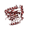
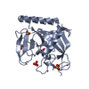
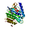
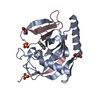
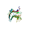
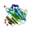
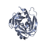
| Title | Crystal structure of Dscam1 Ig7 domain, isoform 5 | ||||||
 Components Components | Down syndrome cell adhesion molecule, isoform AK | ||||||
 Keywords Keywords | CELL ADHESION / Ig fold | ||||||
| Function / homology |  Function and homology information Function and homology informationDSCAM interactions / mushroom body development / detection of molecule of bacterial origin / central nervous system morphogenesis / ventral cord development / detection of mechanical stimulus involved in sensory perception of touch / axon guidance receptor activity / axon extension involved in axon guidance / dendrite self-avoidance / cell-cell adhesion mediator activity ...DSCAM interactions / mushroom body development / detection of molecule of bacterial origin / central nervous system morphogenesis / ventral cord development / detection of mechanical stimulus involved in sensory perception of touch / axon guidance receptor activity / axon extension involved in axon guidance / dendrite self-avoidance / cell-cell adhesion mediator activity / peripheral nervous system development / axonal fasciculation / regulation of axonogenesis / regulation of dendrite morphogenesis / homophilic cell-cell adhesion / antigen binding / neuron development / phagocytosis / axon guidance / central nervous system development / perikaryon / neuron projection / axon / neuronal cell body / dendrite / protein homodimerization activity / extracellular region / identical protein binding / plasma membrane Similarity search - Function | ||||||
| Biological species |  | ||||||
| Method |  X-RAY DIFFRACTION / SYNCHROTRON / MOLECULAR REPLACEMENT / Resolution: 1.948 Å X-RAY DIFFRACTION / SYNCHROTRON / MOLECULAR REPLACEMENT / Resolution: 1.948 Å | ||||||
 Authors Authors | Chen, Q. / Yu, Y. / Li, S. / Cheng, L. | ||||||
 Citation Citation | Journal: Sci Adv / Year: 2016 Title: Structural basis of Dscam1 homodimerization: Insights into context constraint for protein recognition Authors: Li, S.A. / Cheng, L. / Yu, Y. / Chen, Q. | ||||||
| History |
|
- Structure visualization
| Structure viewer | Molecule: MolmilJmol/JSmol |
|---|
- Downloads & links
-Download
| PDBx/mmCIF format | 4wvr.cif.gz | 90.4 KB | Display | PDBx/mmCIF format |
|---|---|---|---|---|
| PDB format | pdb4wvr.ent.gz | 69.7 KB | Display | PDB format |
| PDBx/mmJSON format | 4wvr.json.gz | Tree view | PDBx/mmJSON format | |
| Others |  Other downloads Other downloads |
-Validation report
| Arichive directory | https://data.pdbj.org/pub/pdb/validation_reports/wv/4wvrftp://data.pdbj.org/pub/pdb/validation_reports/wv/4wvr | HTTPS FTP |
|---|
-Related structure data
| Related structure data | 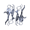 4x5lC  4x83C  4x8xC  4x9bC 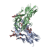 4x9fC 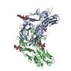 4x9gC 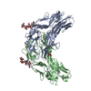 4x9hC 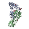 4x9iC  4xb7C  4xb8C  4xhqC  3dmkS S: Starting model for refinement C: citing same article ( |
|---|---|
| Similar structure data |


-Links
 PDBj
PDBj


- Assembly
| Deposited unit | 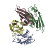
| ||||||||
|---|---|---|---|---|---|---|---|---|---|
| 1 | 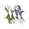
| ||||||||
| 2 | 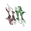
| ||||||||
| Unit cell |
|
-Components
| #1: Protein | Mass: 10708.091 Da / Num. of mol.: 4 / Fragment: UNP residues 617-713 Source method: isolated from a genetically manipulated source Source: (gene. exp.)  #2: Water | ChemComp-HOH / |  Mass: 18.015 Da / Num. of mol.: 296 / Source method: isolated from a natural source / Formula: H2O Mass: 18.015 Da / Num. of mol.: 296 / Source method: isolated from a natural source / Formula: H2OHas protein modification | Y | |
|---|
-Experimental details
-Experiment
| Experiment | Method: X-RAY DIFFRACTION / Number of used crystals: 1 |
|---|
- Sample preparation
| Crystal | Density Matthews: 2.48 Å3/Da / Density % sol: 50.36 % |
|---|---|
| Crystal grow | Temperature: 289 K / Method: vapor diffusion, hanging drop / pH: 6.5 Details: 50mM Calcium chloride dihydrate, 30%(w/v) polyethylene glycol monomethyl ether 550, 0.1M Bis-Tris PH6.5 |
-Data collection
| Diffraction | Mean temperature: 100 K |
|---|---|
| Diffraction source | Source: SYNCHROTRON / Site: BSRF  / Beamline: 3W1A / Wavelength: 1 Å / Beamline: 3W1A / Wavelength: 1 Å |
| Detector | Type: MARRESEARCH / Detector: CCD / Date: Jul 12, 2014 |
| Radiation | Monochromator: Si(111) double-crystal / Protocol: SINGLE WAVELENGTH / Monochromatic (M) / Laue (L): M / Scattering type: x-ray |
| Radiation wavelength | Wavelength: 1 Å / Relative weight: 1 |
| Reflection | Resolution: 1.948→50 Å / Num. obs: 30014 / % possible obs: 99.8 % / Redundancy: 3.8 % / Net I/σ(I): 17.7 |
- Processing
| Software |
| ||||||||||||||||||||||||||||||||||||||||||||||||||||||||||||||||||||||||||||||||||||
|---|---|---|---|---|---|---|---|---|---|---|---|---|---|---|---|---|---|---|---|---|---|---|---|---|---|---|---|---|---|---|---|---|---|---|---|---|---|---|---|---|---|---|---|---|---|---|---|---|---|---|---|---|---|---|---|---|---|---|---|---|---|---|---|---|---|---|---|---|---|---|---|---|---|---|---|---|---|---|---|---|---|---|---|---|---|
| Refinement | Method to determine structure: MOLECULAR REPLACEMENT Starting model: 3DMK Resolution: 1.948→28.912 Å / SU ML: 0.21 / Cross valid method: FREE R-VALUE / σ(F): 1.98 / Phase error: 22.94 / Stereochemistry target values: ML
| ||||||||||||||||||||||||||||||||||||||||||||||||||||||||||||||||||||||||||||||||||||
| Solvent computation | Shrinkage radii: 0.9 Å / VDW probe radii: 1.11 Å / Solvent model: FLAT BULK SOLVENT MODEL | ||||||||||||||||||||||||||||||||||||||||||||||||||||||||||||||||||||||||||||||||||||
| Refinement step | Cycle: LAST / Resolution: 1.948→28.912 Å
| ||||||||||||||||||||||||||||||||||||||||||||||||||||||||||||||||||||||||||||||||||||
| Refine LS restraints |
| ||||||||||||||||||||||||||||||||||||||||||||||||||||||||||||||||||||||||||||||||||||
| LS refinement shell |
|
