 Movie
Movie Controller

[English] 日本語
 Yorodumi
- PDB-4wt5: The C-terminal domain of Rubisco Accumulation Factor 1 from Arabi... -
+ Open data

- Basic information
| Entry | Database: PDB / ID: 4wt5 | ||||||
|---|---|---|---|---|---|---|---|
| Title | The C-terminal domain of Rubisco Accumulation Factor 1 from Arabidopsis thaliana, crystal form II | ||||||
 Components Components | Rubisco Accumulation Factor 1, isoform 2 | ||||||
 Keywords Keywords | CHAPERONE / assembly chaperone | ||||||
| Function / homology |  Function and homology information Function and homology informationribulose bisphosphate carboxylase complex assembly / chloroplast stroma / chloroplast / cytosol Similarity search - Function | ||||||
| Biological species |  | ||||||
| Method |  X-RAY DIFFRACTION / SYNCHROTRON / MOLECULAR REPLACEMENT / molecular replacement / Resolution: 2.568 Å X-RAY DIFFRACTION / SYNCHROTRON / MOLECULAR REPLACEMENT / molecular replacement / Resolution: 2.568 Å | ||||||
 Authors Authors | Hauser, T. / Bhat, J.Y. / Milicic, G. / Wendler, P. / Hartl, F.U. / Bracher, A. / Hayer-Hartl, M. | ||||||
 Citation Citation | Journal: Nat Struct Mol Biol / Year: 2015 Title: Structure and mechanism of the Rubisco-assembly chaperone Raf1. Authors: Thomas Hauser / Javaid Y Bhat / Goran Miličić / Petra Wendler / F Ulrich Hartl / Andreas Bracher / Manajit Hayer-Hartl /  Abstract: Biogenesis of the photosynthetic enzyme Rubisco, a complex of eight large (RbcL) and eight small (RbcS) subunits, requires assembly chaperones. Here we analyzed the role of Rubisco accumulation ...Biogenesis of the photosynthetic enzyme Rubisco, a complex of eight large (RbcL) and eight small (RbcS) subunits, requires assembly chaperones. Here we analyzed the role of Rubisco accumulation factor1 (Raf1), a dimer of ∼40-kDa subunits. We find that Raf1 from Synechococcus elongatus acts downstream of chaperonin-assisted RbcL folding by stabilizing RbcL antiparallel dimers for assembly into RbcL8 complexes with four Raf1 dimers bound. Raf1 displacement by RbcS results in holoenzyme formation. Crystal structures show that Raf1 from Arabidopsis thaliana consists of a β-sheet dimerization domain and a flexibly linked α-helical domain. Chemical cross-linking and EM reconstruction indicate that the β-domains bind along the equator of each RbcL2 unit, and the α-helical domains embrace the top and bottom edges of RbcL2. Raf1 fulfills a role similar to that of the assembly chaperone RbcX, thus suggesting that functionally redundant factors ensure efficient Rubisco biogenesis. | ||||||
| History |
|
- Structure visualization
| Structure viewer | Molecule: MolmilJmol/JSmol |
|---|
- Downloads & links
-Download
| PDBx/mmCIF format | 4wt5.cif.gz | 70.7 KB | Display | PDBx/mmCIF format |
|---|---|---|---|---|
| PDB format | pdb4wt5.ent.gz | 51.2 KB | Display | PDB format |
| PDBx/mmJSON format | 4wt5.json.gz | Tree view | PDBx/mmJSON format | |
| Others |  Other downloads Other downloads |
-Validation report
| Arichive directory | https://data.pdbj.org/pub/pdb/validation_reports/wt/4wt5ftp://data.pdbj.org/pub/pdb/validation_reports/wt/4wt5 | HTTPS FTP |
|---|
-Related structure data
| Related structure data |  3051C  3052C  3053C 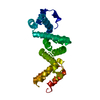 4wt3C  4wt4SC S: Starting model for refinement C: citing same article ( |
|---|---|
| Similar structure data |


-Links
 PDBj
PDBj
- Assembly
| Deposited unit | 
| ||||||||||||||||||
|---|---|---|---|---|---|---|---|---|---|---|---|---|---|---|---|---|---|---|---|
| 1 |
| ||||||||||||||||||
| Unit cell |
| ||||||||||||||||||
| Noncrystallographic symmetry (NCS) | NCS domain:
NCS domain segments: Component-ID: _ / Ens-ID: 1 / Beg auth comp-ID: ILE / Beg label comp-ID: ILE / End auth comp-ID: ASP / End label comp-ID: ASP / Refine code: _ / Auth seq-ID: 288 - 437 / Label seq-ID: 8 - 157
| ||||||||||||||||||
| Details | biological unit is a dimer, a.u. contains one dimer |
-Components
| #1: Protein | Mass: 18839.576 Da / Num. of mol.: 2 / Fragment: Raf1 beta-domain, UNP residues 281-449 Source method: isolated from a genetically manipulated source Source: (gene. exp.)  #2: Water | ChemComp-HOH / |  Mass: 18.015 Da / Num. of mol.: 9 / Source method: isolated from a natural source / Formula: H2O Mass: 18.015 Da / Num. of mol.: 9 / Source method: isolated from a natural source / Formula: H2O |
|---|
-Experimental details
-Experiment
| Experiment | Method: X-RAY DIFFRACTION / Number of used crystals: 1 |
|---|
- Sample preparation
| Crystal | Density Matthews: 2.29 Å3/Da / Density % sol: 46.36 % |
|---|---|
| Crystal grow | Temperature: 291 K / Method: vapor diffusion, sitting drop / pH: 8 / Details: 200 mM KH2PO4 and 20% (w/v) PEG-5000MME |
-Data collection
| Diffraction | Mean temperature: 100 K | ||||||||||||||||||||||||||||||||||||||||||||||||||||||||||||||||||||||||||||||||||||||||||||||||||||||||||||||
|---|---|---|---|---|---|---|---|---|---|---|---|---|---|---|---|---|---|---|---|---|---|---|---|---|---|---|---|---|---|---|---|---|---|---|---|---|---|---|---|---|---|---|---|---|---|---|---|---|---|---|---|---|---|---|---|---|---|---|---|---|---|---|---|---|---|---|---|---|---|---|---|---|---|---|---|---|---|---|---|---|---|---|---|---|---|---|---|---|---|---|---|---|---|---|---|---|---|---|---|---|---|---|---|---|---|---|---|---|---|---|---|
| Diffraction source | Source: SYNCHROTRON / Site: SLS  / Beamline: X10SA / Wavelength: 0.97856 Å / Beamline: X10SA / Wavelength: 0.97856 Å | ||||||||||||||||||||||||||||||||||||||||||||||||||||||||||||||||||||||||||||||||||||||||||||||||||||||||||||||
| Detector | Type: DECTRIS PILATUS 6M / Detector: PIXEL / Date: May 9, 2013 | ||||||||||||||||||||||||||||||||||||||||||||||||||||||||||||||||||||||||||||||||||||||||||||||||||||||||||||||
| Radiation | Protocol: SINGLE WAVELENGTH / Monochromatic (M) / Laue (L): M / Scattering type: x-ray | ||||||||||||||||||||||||||||||||||||||||||||||||||||||||||||||||||||||||||||||||||||||||||||||||||||||||||||||
| Radiation wavelength | Wavelength: 0.97856 Å / Relative weight: 1 | ||||||||||||||||||||||||||||||||||||||||||||||||||||||||||||||||||||||||||||||||||||||||||||||||||||||||||||||
| Reflection | Resolution: 2.568→71.633 Å / Num. all: 11314 / Num. obs: 11314 / % possible obs: 96.7 % / Redundancy: 4.3 % / Rpim(I) all: 0.051 / Rrim(I) all: 0.11 / Rsym value: 0.097 / Net I/av σ(I): 5.931 / Net I/σ(I): 11.9 / Num. measured all: 48923 | ||||||||||||||||||||||||||||||||||||||||||||||||||||||||||||||||||||||||||||||||||||||||||||||||||||||||||||||
| Reflection shell | Diffraction-ID: 1 / Rejects: _
|
-Phasing
| Phasing | Method: molecular replacement | |||||||||
|---|---|---|---|---|---|---|---|---|---|---|
| Phasing MR |
|
- Processing
| Software |
| |||||||||||||||||||||||||||||||||||||||||||||
|---|---|---|---|---|---|---|---|---|---|---|---|---|---|---|---|---|---|---|---|---|---|---|---|---|---|---|---|---|---|---|---|---|---|---|---|---|---|---|---|---|---|---|---|---|---|---|
| Refinement | Method to determine structure: MOLECULAR REPLACEMENT Starting model: 4WT4 Resolution: 2.568→30 Å / Cor.coef. Fo:Fc: 0.949 / Cor.coef. Fo:Fc free: 0.914 / WRfactor Rfree: 0.2643 / WRfactor Rwork: 0.1927 / FOM work R set: 0.7932 / SU B: 12.968 / SU ML: 0.271 / SU R Cruickshank DPI: 0.6741 / SU Rfree: 0.3411 / Cross valid method: THROUGHOUT / σ(F): 0 / ESU R: 0.674 / ESU R Free: 0.341 / Stereochemistry target values: MAXIMUM LIKELIHOOD Details: HYDROGENS HAVE BEEN USED IF PRESENT IN THE INPUT U VALUES : REFINED INDIVIDUALLY
| |||||||||||||||||||||||||||||||||||||||||||||
| Solvent computation | Ion probe radii: 0.8 Å / Shrinkage radii: 0.8 Å / VDW probe radii: 1.2 Å / Solvent model: MASK | |||||||||||||||||||||||||||||||||||||||||||||
| Displacement parameters | Biso max: 126.78 Å2 / Biso mean: 59.375 Å2 / Biso min: 26.46 Å2
| |||||||||||||||||||||||||||||||||||||||||||||
| Refinement step | Cycle: final / Resolution: 2.568→30 Å
| |||||||||||||||||||||||||||||||||||||||||||||
| Refine LS restraints |
| |||||||||||||||||||||||||||||||||||||||||||||
| Refine LS restraints NCS | Ens-ID: 1 / Number: 120 / Refine-ID: X-RAY DIFFRACTION / Type: interatomic distance / Rms dev position: 0.21 Å / Weight position: 0.05
| |||||||||||||||||||||||||||||||||||||||||||||
| LS refinement shell | Resolution: 2.568→2.634 Å / Total num. of bins used: 20
|
