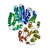
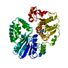
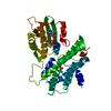
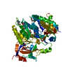
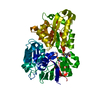
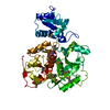
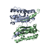
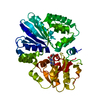
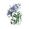
Entry Database : PDB / ID : 4v3cTitle The structure of alpha2,3-sialyltransferase variant 2 from Pasteurella dagmatis in complex with the donor product CMP SIALYLTRANSFERASE Keywords Function / homology Function Domain/homology Component
/ / / / / / / Biological species PASTEURELLA DAGMATIS (bacteria)Method / / / Resolution : 1.84 Å Authors Pavkov-Keller, T. / Schmoelzer, K. / Czabany, T. / Luley-Goedl, C. / Ribitsch, D. / Schwab, H. / Nidetzky, B. / Gruber, K. Journal : Chem.Commun.(Camb.) / Year : 2015Title : Complete Switch from Alpha2,3- to Alpha2,6-Regioselectivity in Pasteurella Dagmatis Beta-D-Galactoside Sialyltransferase by Active-Site RedesignAuthors : Schmoelzer, K. / Czabany, T. / Pavkov-Keller, T. / Luley-Goedl, C. / Ribitsch, D. / Schwab, H. / Gruber, K. / Nidetzky, B. History Deposition Oct 17, 2014 Deposition site / Processing site Revision 1.0 Apr 8, 2015 Provider / Type Revision 1.1 Sep 13, 2017 Group / Category / Item Revision 1.2 Jan 10, 2024 Group Data collection / Database references ... Data collection / Database references / Derived calculations / Other / Refinement description Category chem_comp_atom / chem_comp_bond ... chem_comp_atom / chem_comp_bond / database_2 / pdbx_database_status / pdbx_initial_refinement_model / struct_site Item _database_2.pdbx_DOI / _database_2.pdbx_database_accession ... _database_2.pdbx_DOI / _database_2.pdbx_database_accession / _pdbx_database_status.status_code_sf / _struct_site.pdbx_auth_asym_id / _struct_site.pdbx_auth_comp_id / _struct_site.pdbx_auth_seq_id Revision 2.0 Dec 17, 2025 Group Atomic model / Data collection ... Atomic model / Data collection / Derived calculations / Non-polymer description / Structure summary Category atom_site / chem_comp ... atom_site / chem_comp / chem_comp_atom / chem_comp_bond / pdbx_entity_nonpoly / pdbx_entry_details / pdbx_nonpoly_scheme / struct_site Item _atom_site.B_iso_or_equiv / _atom_site.Cartn_x ... _atom_site.B_iso_or_equiv / _atom_site.Cartn_x / _atom_site.Cartn_y / _atom_site.Cartn_z / _atom_site.auth_atom_id / _atom_site.auth_comp_id / _atom_site.label_atom_id / _atom_site.label_comp_id / _atom_site.type_symbol / _chem_comp.id / _chem_comp.mon_nstd_flag / _chem_comp.type / _chem_comp_atom.atom_id / _chem_comp_atom.comp_id / _chem_comp_atom.type_symbol / _chem_comp_bond.atom_id_1 / _chem_comp_bond.atom_id_2 / _chem_comp_bond.comp_id / _chem_comp_bond.value_order / _pdbx_entity_nonpoly.comp_id / _pdbx_nonpoly_scheme.mon_id / _pdbx_nonpoly_scheme.pdb_mon_id / _struct_site.details / _struct_site.pdbx_auth_comp_id
Show all Show less
 Movie
Movie Controller

 Yorodumi
 Open data
 Basic information
 Components
 Keywords
 Function and homology information
 PASTEURELLA DAGMATIS (bacteria)
 X-RAY DIFFRACTION /
  Authors
 Citation
 Structure visualization
 Downloads & links
 Other downloads


 PDBj
PDBj Assembly
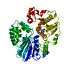

 Mass: 323.197 Da / Num. of mol.: 1 / Source method: obtained synthetically / Formula: C9H14N3O8P
 Mass: 18.015 Da / Num. of mol.: 297 / Source method: isolated from a natural source / Formula: H2O
 Sample preparation
 / Beamline: ID29 / Wavelength: 0.9791
  Processing