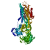
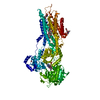
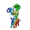
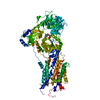
[English] 日本語
Yorodumi
- PDB-4uu1: CRYSTAL STRUCTURE OF (SR) CALCIUM-ATPASE E2(TG) IN THE PRESENCE O... -

+
Open data


ID or keywords:

Loading...

-
Basic information

Entry
Database: PDB / ID: 4uu1
TitleCRYSTAL STRUCTURE OF (SR) CALCIUM-ATPASE E2(TG) IN THE PRESENCE OF DOPC
ComponentsSARCOPLASMIC ENDOPLASMIC RETICULUM CALCIUM ATPASE
KeywordsHYDROLASE / CA2+-ATPASE / P-TYPE ATPASE / CATION PUMP / MEMBRANE PROTEIN / LIPID BINDING / LIPID BINDING SITES / DOPC
Function / homology
Function and homology information


positive regulation of cardiac muscle cell contraction / positive regulation of calcium ion import into sarcoplasmic reticulum / H zone / positive regulation of fast-twitch skeletal muscle fiber contraction / calcium ion import into sarcoplasmic reticulum / negative regulation of striated muscle contraction / regulation of striated muscle contraction / positive regulation of ATPase-coupled calcium transmembrane transporter activity / P-type Ca2+ transporter / P-type calcium transporter activity ...positive regulation of cardiac muscle cell contraction / positive regulation of calcium ion import into sarcoplasmic reticulum / H zone / positive regulation of fast-twitch skeletal muscle fiber contraction / calcium ion import into sarcoplasmic reticulum / negative regulation of striated muscle contraction / regulation of striated muscle contraction / positive regulation of ATPase-coupled calcium transmembrane transporter activity / P-type Ca2+ transporter / P-type calcium transporter activity / I band / endoplasmic reticulum-Golgi intermediate compartment / sarcoplasmic reticulum membrane / sarcoplasmic reticulum / intracellular calcium ion homeostasis / calcium ion transport / calcium ion binding / endoplasmic reticulum membrane / perinuclear region of cytoplasm / endoplasmic reticulum / ATP hydrolysis activity / ATP binding / membrane / metal ion binding
Similarity search - Function
Calcium-transporting ATPase, transmembrane domain / Calcium-transporting ATPase, transmembrane domain / P-type ATPase, subfamily IIA, SERCA-type / Calcium-transporting ATPase, cytoplasmic transduction domain A / Calcium-transporting ATPase, cytoplasmic transduction domain A / Calcium-transporting ATPase, cytoplasmic domain N / Calcium-transporting ATPase, cytoplasmic domain N / haloacid dehalogenase-like hydrolase / Cation-transporting P-type ATPase, C-terminal / Cation transporting ATPase, C-terminus ...Calcium-transporting ATPase, transmembrane domain / Calcium-transporting ATPase, transmembrane domain / P-type ATPase, subfamily IIA, SERCA-type / Calcium-transporting ATPase, cytoplasmic transduction domain A / Calcium-transporting ATPase, cytoplasmic transduction domain A / Calcium-transporting ATPase, cytoplasmic domain N / Calcium-transporting ATPase, cytoplasmic domain N / haloacid dehalogenase-like hydrolase / Cation-transporting P-type ATPase, C-terminal / Cation transporting ATPase, C-terminus / Cation transporter/ATPase, N-terminus / Cation-transporting P-type ATPase, N-terminal / Cation transporter/ATPase, N-terminus / Cation transport ATPase (P-type) / E1-E2 ATPase / P-type ATPase, haloacid dehalogenase domain / P-type ATPase, phosphorylation site / P-type ATPase, cytoplasmic domain N / E1-E2 ATPases phosphorylation site. / P-type ATPase, A domain superfamily / P-type ATPase / P-type ATPase, transmembrane domain superfamily / HAD superfamily/HAD-like / haloacid dehalogenase-like hydrolase / HAD superfamily / HAD-like superfamily / Distorted Sandwich / Up-down Bundle / Rossmann fold / 3-Layer(aba) Sandwich / Mainly Beta / Mainly Alpha / Alpha Beta
Similarity search - Domain/homology
PHOSPHOMETHYLPHOSPHONIC ACID ADENYLATE ESTER / : / 1,2-DIOLEOYL-SN-GLYCERO-3-PHOSPHOCHOLINE / Chem-TG1 / Calcium-transporting ATPase / Sarcoplasmic/endoplasmic reticulum calcium ATPase 1
Similarity search - Component
Biological speciesORYCTOLAGUS CUNICULUS (rabbit)
MethodX-RAY DIFFRACTION / SYNCHROTRON / MOLECULAR REPLACEMENT / Resolution: 2.8 Å
AuthorsDrachmann, N.D. / Olesen, C. / Moeller, J.V. / Guo, Z. / Nissen, P. / Bublitz, M.
CitationJournal: FEBS J. / Year: 2014
Title: Comparing Crystal Structures of Ca(2+) -ATPase in the Presence of Different Lipids.
Authors: Drachmann, N.D. / Olesen, C. / Moller, J.V. / Guo, Z. / Nissen, P. / Bublitz, M.
History
DepositionJul 24, 2014Deposition site: PDBE / Processing site: PDBE
Revision 1.0Oct 1, 2014Provider: repository / Type: Initial release
Revision 1.1Oct 23, 2019Group: Data collection / Database references ...Data collection / Database references / Derived calculations / Other
Category: pdbx_database_status / struct_conn / struct_ref_seq_dif
Item: _pdbx_database_status.status_code_sf / _struct_conn.pdbx_leaving_atom_flag / _struct_ref_seq_dif.details
Revision 1.2Jan 10, 2024Group: Data collection / Database references ...Data collection / Database references / Derived calculations / Refinement description / Structure summary
Category: chem_comp / chem_comp_atom ...chem_comp / chem_comp_atom / chem_comp_bond / database_2 / entity / pdbx_entity_nonpoly / pdbx_initial_refinement_model / pdbx_struct_conn_angle / struct_conn / struct_site
Item: _chem_comp.name / _database_2.pdbx_DOI ..._chem_comp.name / _database_2.pdbx_DOI / _database_2.pdbx_database_accession / _entity.pdbx_description / _pdbx_entity_nonpoly.name / _pdbx_struct_conn_angle.ptnr1_auth_comp_id / _pdbx_struct_conn_angle.ptnr1_auth_seq_id / _pdbx_struct_conn_angle.ptnr1_label_asym_id / _pdbx_struct_conn_angle.ptnr1_label_atom_id / _pdbx_struct_conn_angle.ptnr1_label_comp_id / _pdbx_struct_conn_angle.ptnr1_label_seq_id / _pdbx_struct_conn_angle.ptnr2_auth_comp_id / _pdbx_struct_conn_angle.ptnr2_auth_seq_id / _pdbx_struct_conn_angle.ptnr2_label_asym_id / _pdbx_struct_conn_angle.ptnr2_label_atom_id / _pdbx_struct_conn_angle.ptnr2_label_comp_id / _pdbx_struct_conn_angle.ptnr3_auth_comp_id / _pdbx_struct_conn_angle.ptnr3_auth_seq_id / _pdbx_struct_conn_angle.ptnr3_label_asym_id / _pdbx_struct_conn_angle.ptnr3_label_atom_id / _pdbx_struct_conn_angle.ptnr3_label_comp_id / _pdbx_struct_conn_angle.ptnr3_label_seq_id / _pdbx_struct_conn_angle.value / _struct_conn.pdbx_dist_value / _struct_conn.ptnr1_auth_comp_id / _struct_conn.ptnr1_auth_seq_id / _struct_conn.ptnr1_label_asym_id / _struct_conn.ptnr1_label_atom_id / _struct_conn.ptnr1_label_comp_id / _struct_conn.ptnr1_label_seq_id / _struct_conn.ptnr2_auth_comp_id / _struct_conn.ptnr2_auth_seq_id / _struct_conn.ptnr2_label_asym_id / _struct_conn.ptnr2_label_atom_id / _struct_conn.ptnr2_label_comp_id / _struct_conn.ptnr2_label_seq_id / _struct_site.pdbx_auth_asym_id / _struct_site.pdbx_auth_comp_id / _struct_site.pdbx_auth_seq_id

-
Structure visualization

Structure viewerMolecule:
MolmilJmol/JSmol

Downloads & links

-
Assembly

Deposited unit
A: SARCOPLASMIC ENDOPLASMIC RETICULUM CALCIUM ATPASE
hetero molecules


Theoretical massNumber of molelcules
Total (without water)113,39410
Polymers109,6291
Non-polymers3,7659
Water00
1


  • Idetical with deposited unit
  • defined by author&software
TypeNameSymmetry operationNumber
identity operation1_555x,y,z1
MethodPISA
Unit cell
Length a, b, c (Å)71.703, 71.703, 591.318
Angle α, β, γ (deg.)90.00, 90.00, 90.00
Int Tables number92
Space group name H-MP41212

-
Components

-
Protein , 1 types, 1 molecules A

#1: Protein SARCOPLASMIC ENDOPLASMIC RETICULUM CALCIUM ATPASE


Mass: 109628.617 Da / Num. of mol.: 1 / Source method: isolated from a natural source / Source: (natural) ORYCTOLAGUS CUNICULUS (rabbit) / Organ: HIND LEG MUSCLE / Tissue: FAST TWITCH SKELETAL MUSCLE
References: UniProt: B6CAM1, UniProt: P04191*PLUS, EC: 3.6.3.8

-
Non-polymers , 6 types, 9 molecules

#2: Chemical ChemComp-TG1 / OCTANOIC ACID [3S-[3ALPHA, 3ABETA, 4ALPHA, 6BETA, 6ABETA, 7BETA, 8ALPHA(Z), 9BALPHA]]-6-(ACETYLOXY)-2,3,-3A,4,5,6,6A,7,8,9B-DECAHYDRO-3,3A-DIHYDROXY-3,6,9-TRIMETHYL-8-[(2-METHYL-1-OXO-2-BUTENYL)OX Y]-2-OXO-4-(1-OXOBUTOXY)-AZULENO[4,5-B]FURAN-7-YL ESTER / THAPSIGARGIN


Mass: 650.754 Da / Num. of mol.: 1 / Source method: obtained synthetically / Formula: C34H50O12 / Comment: inhibitor*YM
#3: Chemical ChemComp-PCW / 1,2-DIOLEOYL-SN-GLYCERO-3-PHOSPHOCHOLINE / (Z,Z)-4-HYDROXY-N,N,N-TRIMETHYL-10-OXO-7-[(1-OXO-9-OCTADECENYL)OXY]-3,5,9-TRIOXA-4-PHOSPHAHEPTACOS-18-EN-1-AMINIUM-4-OXIDE


Mass: 787.121 Da / Num. of mol.: 3 / Source method: obtained synthetically / Formula: C44H85NO8P / Comment: DOPC, phospholipid*YM
#4: Chemical ChemComp-GOL / GLYCEROL / GLYCERIN / PROPANE-1,2,3-TRIOL


Mass: 92.094 Da / Num. of mol.: 2 / Source method: obtained synthetically / Formula: C3H8O3
#5: Chemical ChemComp-ACP / PHOSPHOMETHYLPHOSPHONIC ACID ADENYLATE ESTER / ADENOSINE-5'-[BETA, GAMMA-METHYLENE]TRIPHOSPHATE


Mass: 505.208 Da / Num. of mol.: 1 / Source method: obtained synthetically / Formula: C11H18N5O12P3 / Comment: AMP-PCP, energy-carrying molecule analogue*YM
#6: Chemical ChemComp-K / POTASSIUM ION


Mass: 39.098 Da / Num. of mol.: 1 / Source method: obtained synthetically / Formula: K
#7: Chemical ChemComp-MG / MAGNESIUM ION


Mass: 24.305 Da / Num. of mol.: 1 / Source method: obtained synthetically / Formula: Mg

-
Details

Nonpolymer details1,2-DIOLEOYL-SN-GLYCERO-3-PHOSPHOCHOLINE (PCW): PARTIALLY MODELED LIGAND

-
Experimental details

-
Experiment

ExperimentMethod: X-RAY DIFFRACTION / Number of used crystals: 1

-
Sample preparation

CrystalDensity Matthews: 3.36 Å3/Da / Density % sol: 63.38 % / Description: NONE
Crystal growpH: 6.8
Details: 18 % (V/V) PEG2000, 100 MM MGSO4, 3 % (V/V) TERT-BUOH, pH 6.8

-
Data collection

DiffractionMean temperature: 100 K
Diffraction sourceSource: SYNCHROTRON / Site: SLS / Beamline: X06SA / Wavelength: 0.97935
DetectorType: DECTRIS PILATUS 6M / Detector: PIXEL / Date: Feb 10, 2012
RadiationMonochromator: LN2 COOLED FIXED-EXIT SI(111) MONOCHROMATOR / Protocol: SINGLE WAVELENGTH / Monochromatic (M) / Laue (L): M / Scattering type: x-ray
Radiation wavelengthWavelength: 0.97935 Å / Relative weight: 1
ReflectionResolution: 2.8→73.9 Å / Num. obs: 39919 / % possible obs: 100 % / Observed criterion σ(I): -3 / Redundancy: 13.3 % / Biso Wilson estimate: 79.73 Å2 / Rmerge(I) obs: 0.09 / Net I/σ(I): 17.06
Reflection shellResolution: 2.8→2.95 Å / Redundancy: 13.3 % / Rmerge(I) obs: 1.21 / Mean I/σ(I) obs: 2.1 / % possible all: 100

-
Processing

Software
NameVersionClassification
PHENIX(PHENIX.REFINE)refinement
XDSdata reduction
XSCALEdata scaling
PHASERphasing
RefinementMethod to determine structure: MOLECULAR REPLACEMENT
Starting model: PDB ENTRY 2C8K
Resolution: 2.8→73.915 Å / SU ML: 0.45 / σ(F): 2 / Phase error: 30.13 / Stereochemistry target values: ML
RfactorNum. reflection% reflection
Rfree0.2929 1995 5 %
Rwork0.2211 --
obs0.2246 39916 99.97 %
Solvent computationShrinkage radii: 0.9 Å / VDW probe radii: 1.11 Å / Solvent model: FLAT BULK SOLVENT MODEL
Refinement stepCycle: LAST / Resolution: 2.8→73.915 Å
ProteinNucleic acidLigandSolventTotal
Num. atoms7674 0 160 0 7834
Refine LS restraints
Refine-IDTypeDev idealNumber
X-RAY DIFFRACTIONf_bond_d0.0117984
X-RAY DIFFRACTIONf_angle_d1.18110822
X-RAY DIFFRACTIONf_dihedral_angle_d17.3133059
X-RAY DIFFRACTIONf_chiral_restr0.0451240
X-RAY DIFFRACTIONf_plane_restr0.0051365
LS refinement shell
Resolution (Å)Rfactor RfreeNum. reflection RfreeRfactor RworkNum. reflection RworkRefine-ID% reflection obs (%)
2.8-2.870.33471390.26852667X-RAY DIFFRACTION100
2.87-2.94760.31561300.26812601X-RAY DIFFRACTION100
2.9476-3.03440.31581410.26432622X-RAY DIFFRACTION100
3.0344-3.13230.3911430.27092670X-RAY DIFFRACTION100
3.1323-3.24430.32721410.26312646X-RAY DIFFRACTION100
3.2443-3.37420.32761410.25722654X-RAY DIFFRACTION100
3.3742-3.52770.34951370.25052650X-RAY DIFFRACTION100
3.5277-3.71370.34411410.25822681X-RAY DIFFRACTION100
3.7137-3.94640.31641420.24392686X-RAY DIFFRACTION100
3.9464-4.2510.30831440.20892725X-RAY DIFFRACTION100
4.251-4.67880.25631410.17882707X-RAY DIFFRACTION100
4.6788-5.35560.26131490.18472755X-RAY DIFFRACTION100
5.3556-6.74680.29571460.23212823X-RAY DIFFRACTION100
6.7468-73.94050.25631600.20513034X-RAY DIFFRACTION100
Refinement TLS params.

Method: refined / Refine-ID: X-RAY DIFFRACTION

IDL112)L122)L132)L222)L232)L332)S11 (Å °)S12 (Å °)S13 (Å °)S21 (Å °)S22 (Å °)S23 (Å °)S31 (Å °)S32 (Å °)S33 (Å °)T112)T122)T132)T222)T232)T332)Origin x (Å)Origin y (Å)Origin z (Å)
14.8223-1.4688-0.5083.78531.2488.16340.2536-0.2758-0.6251-0.13040.124-0.49770.6270.1119-0.26220.50580.0228-0.18640.39810.12840.7942-1.45476.9257-17.3722
21.4201-0.17252.53270.5499-0.65075.5985-0.05890.2887-0.2212-0.43450.14840.01790.4224-0.0381-0.13281.1613-0.1109-0.02970.6662-0.05570.5164-15.044125.9276-63.9982
35.42220.0674-0.69920.81541.57875.4286-0.2717-0.4376-0.186-0.11990.00730.2863-0.4719-0.2950.22911.01210.021-0.21810.51890.13660.5893-10.391539.1079-27.4121
43.42470.843-0.20635.51460.23514.5907-0.12020.25110.06320.4908-0.0666-0.6469-0.49490.61650.15970.8468-0.1408-0.22010.57640.10850.4319.907638.9642-0.7939
51.7286-0.1561.07710.00330.05333.0672-0.3350.43750.1824-0.01430.0684-0.0189-1.01470.07410.26051.58590.0344-0.08950.80540.1290.5115-14.566146.7768-73.9492
Refinement TLS group
IDRefine-IDRefine TLS-IDSelection details
1X-RAY DIFFRACTION1CHAIN A AND (RESID 1:43 OR RESID 123:228)
2X-RAY DIFFRACTION2CHAIN A AND (RESID 44:122 OR RESID 229:329)
3X-RAY DIFFRACTION3CHAIN A AND (RESID 330:359 OR RESID 605:745 OR RESID 1301)
4X-RAY DIFFRACTION4CHAIN A AND RESID 360:604
5X-RAY DIFFRACTION5CHAIN A AND RESID 746:994

+
About Yorodumi

-
News

-
Feb 9, 2022. New format data for meta-information of EMDB entries

New format data for meta-information of EMDB entries

  • Version 3 of the EMDB header file is now the official format.
  • The previous official version 1.9 will be removed from the archive.

Related info.:EMDB header

External links:wwPDB to switch to version 3 of the EMDB data model

-
Aug 12, 2020. Covid-19 info

Covid-19 info

URL: https://pdbjlvh1.pdbj.org/emnavi/covid19.php

New page: Covid-19 featured information page in EM Navigator.

Related info.:Covid-19 info / Mar 5, 2020. Novel coronavirus structure data

+
Mar 5, 2020. Novel coronavirus structure data

Novel coronavirus structure data

Related info.:Yorodumi Speices / Aug 12, 2020. Covid-19 info

External links:COVID-19 featured content - PDBj / Molecule of the Month (242):Coronavirus Proteases

+
Jan 31, 2019. EMDB accession codes are about to change! (news from PDBe EMDB page)

EMDB accession codes are about to change! (news from PDBe EMDB page)

  • The allocation of 4 digits for EMDB accession codes will soon come to an end. Whilst these codes will remain in use, new EMDB accession codes will include an additional digit and will expand incrementally as the available range of codes is exhausted. The current 4-digit format prefixed with “EMD-” (i.e. EMD-XXXX) will advance to a 5-digit format (i.e. EMD-XXXXX), and so on. It is currently estimated that the 4-digit codes will be depleted around Spring 2019, at which point the 5-digit format will come into force.
  • The EM Navigator/Yorodumi systems omit the EMD- prefix.

Related info.:Q: What is EMD? / ID/Accession-code notation in Yorodumi/EM Navigator

External links:EMDB Accession Codes are Changing Soon! / Contact to PDBj

+
Jul 12, 2017. Major update of PDB

Major update of PDB

  • wwPDB released updated PDB data conforming to the new PDBx/mmCIF dictionary.
  • This is a major update changing the version number from 4 to 5, and with Remediation, in which all the entries are updated.
  • In this update, many items about electron microscopy experimental information are reorganized (e.g. em_software).
  • Now, EM Navigator and Yorodumi are based on the updated data.

External links:wwPDB Remediation / Enriched Model Files Conforming to OneDep Data Standards Now Available in the PDB FTP Archive

-
Yorodumi

Thousand views of thousand structures

  • Yorodumi is a browser for structure data from EMDB, PDB, SASBDB, etc.
  • This page is also the successor to EM Navigator detail page, and also detail information page/front-end page for Omokage search.
  • The word "yorodu" (or yorozu) is an old Japanese word meaning "ten thousand". "mi" (miru) is to see.

Related info.:EMDB / PDB / SASBDB / Comparison of 3 databanks / Yorodumi Search / Aug 31, 2016. New EM Navigator & Yorodumi / Yorodumi Papers / Jmol/JSmol / Function and homology information / Changes in new EM Navigator and Yorodumi

Read more