 Movie
Movie Controller

[English] 日本語
 Yorodumi
- PDB-4usz: Crystal structure of the first bacterial vanadium dependant iodop... -
+ Open data

- Basic information
| Entry | Database: PDB / ID: 4usz | ||||||
|---|---|---|---|---|---|---|---|
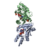
| Title | Crystal structure of the first bacterial vanadium dependant iodoperoxidase | ||||||
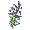
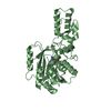
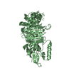
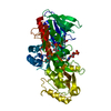
 Components Components | VANADIUM-DEPENDENT HALOPEROXIDASE | ||||||
 Keywords Keywords | OXIDOREDUCTASE / MARINE BACTERIUM | ||||||
| Function / homology |  Function and homology information Function and homology informationOxidoreductases; Acting on a peroxide as acceptor; Peroxidases / peroxidase activity Similarity search - Function | ||||||
| Biological species |  ZOBELLIA GALACTANIVORANS (bacteria) ZOBELLIA GALACTANIVORANS (bacteria) | ||||||
| Method |  X-RAY DIFFRACTION / SYNCHROTRON / MOLECULAR REPLACEMENT / Resolution: 2 Å X-RAY DIFFRACTION / SYNCHROTRON / MOLECULAR REPLACEMENT / Resolution: 2 Å | ||||||
 Authors Authors | Rebuffet, E. / Delage, L. / Fournier, J.B. / Rzonca, J. / Potin, P. / Michel, G. / Czjzek, M. / Leblanc, C. | ||||||
 Citation Citation | Journal: Appl.Environ.Microbiol. / Year: 2014 Title: The Bacterial Vanadium Iodoperoxidase from the Marine Flavobacteriaceae Zobellia Galactanivorans Reveals Novel Molecular and Evolutionary Features of Halide Specificity in This Enzyme Family. Authors: Fournier, J.B. / Rebuffet, E. / Delage, L. / Grijol, R. / Meslet-Cladiere, L. / Rzonca, J. / Potin, P. / Michel, G. / Czjzek, M. / Leblanc, C. | ||||||
| History |
|
- Structure visualization
| Structure viewer | Molecule: MolmilJmol/JSmol |
|---|
- Downloads & links
-Download
| PDBx/mmCIF format | 4usz.cif.gz | 102.1 KB | Display | PDBx/mmCIF format |
|---|---|---|---|---|
| PDB format | pdb4usz.ent.gz | 76.5 KB | Display | PDB format |
| PDBx/mmJSON format | 4usz.json.gz | Tree view | PDBx/mmJSON format | |
| Others |  Other downloads Other downloads |
-Validation report
| Arichive directory | https://data.pdbj.org/pub/pdb/validation_reports/us/4uszftp://data.pdbj.org/pub/pdb/validation_reports/us/4usz | HTTPS FTP |
|---|
-Related structure data
| Related structure data | 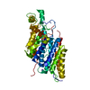 4citSC S: Starting model for refinement C: citing same article ( |
|---|---|
| Similar structure data |


-Links
 PDBj
PDBj


- Assembly
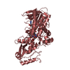
| Deposited unit | 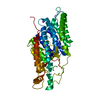
| ||||||||
|---|---|---|---|---|---|---|---|---|---|
| 1 |
| ||||||||
| Unit cell |
|
-Components
| #1: Protein | Mass: 51495.078 Da / Num. of mol.: 1 Source method: isolated from a genetically manipulated source Details: VANADATE COFACTOR LINKED THROUGH HIS 416 / Source: (gene. exp.) ZOBELLIA GALACTANIVORANS (bacteria)Description: GERMAN COLLECTION OF MICROORGANISMS (DSM) AND AVAILABLE AT STATION BIOLOGIQUE DE ROSCOFF, FRANCE Production host: References: UniProt: G0LAH5, EC: 1.11.1.8, Oxidoreductases; Acting on a peroxide as acceptor; Peroxidases |
|---|---|
| #2: Chemical | ChemComp-VO4 /   Mass: 114.939 Da / Num. of mol.: 1 / Source method: obtained synthetically / Formula: VO4 Mass: 114.939 Da / Num. of mol.: 1 / Source method: obtained synthetically / Formula: VO4 |
| #3: Chemical | ChemComp-NA /   Mass: 22.990 Da / Num. of mol.: 1 / Source method: obtained synthetically / Formula: Na Mass: 22.990 Da / Num. of mol.: 1 / Source method: obtained synthetically / Formula: Na |
| #4: Water | ChemComp-HOH /  Mass: 18.015 Da / Num. of mol.: 283 / Source method: isolated from a natural source / Formula: H2O Mass: 18.015 Da / Num. of mol.: 283 / Source method: isolated from a natural source / Formula: H2O |
-Experimental details
-Experiment
| Experiment | Method: X-RAY DIFFRACTION / Number of used crystals: 1 |
|---|
- Sample preparation
| Crystal | Density Matthews: 2.19 Å3/Da / Density % sol: 44 % Description: THIS IS THE NATIVE DATA, WHILE THE STRUCTURE 4CIT CONCERNS THE SE-MET LABELED PROTEIN |
|---|---|
| Crystal grow | pH: 7.2 Details: DROPS: 1 MICROL OF ENZYME AND % PEG 1150, 100 MM PHOSPHATE/CITRATE PH 4.2 AND 2 % GLYCEROL |
-Data collection
| Diffraction | Mean temperature: 100 K |
|---|---|
| Diffraction source | Source: SYNCHROTRON / Site: ESRF  / Beamline: BM30A / Wavelength: 1.04 / Beamline: BM30A / Wavelength: 1.04 |
| Detector | Type: ADSC QUANTUM 315r / Detector: CCD / Date: Nov 24, 2008 |
| Radiation | Protocol: SINGLE WAVELENGTH / Monochromatic (M) / Laue (L): M / Scattering type: x-ray |
| Radiation wavelength | Wavelength: 1.04 Å / Relative weight: 1 |
| Reflection | Resolution: 2→69.1 Å / Num. obs: 34478 / % possible obs: 99.6 % / Observed criterion σ(I): 0.1 / Redundancy: 3.8 % / Rmerge(I) obs: 0.1 / Net I/σ(I): 12.4 |
| Reflection shell | Resolution: 2→2.06 Å / Redundancy: 3.9 % / Rmerge(I) obs: 0.31 / Mean I/σ(I) obs: 4.56 / % possible all: 99.9 |
- Processing
| Software |
| ||||||||||||||||||||||||||||||||||||||||||||||||||||||||||||||||||||||||||||||||||||||||||||||||||||||||||||||||||||||||||||||||||||||||||||||||||||||||||||||||||||||||||||||||||||||
|---|---|---|---|---|---|---|---|---|---|---|---|---|---|---|---|---|---|---|---|---|---|---|---|---|---|---|---|---|---|---|---|---|---|---|---|---|---|---|---|---|---|---|---|---|---|---|---|---|---|---|---|---|---|---|---|---|---|---|---|---|---|---|---|---|---|---|---|---|---|---|---|---|---|---|---|---|---|---|---|---|---|---|---|---|---|---|---|---|---|---|---|---|---|---|---|---|---|---|---|---|---|---|---|---|---|---|---|---|---|---|---|---|---|---|---|---|---|---|---|---|---|---|---|---|---|---|---|---|---|---|---|---|---|---|---|---|---|---|---|---|---|---|---|---|---|---|---|---|---|---|---|---|---|---|---|---|---|---|---|---|---|---|---|---|---|---|---|---|---|---|---|---|---|---|---|---|---|---|---|---|---|---|---|
| Refinement | Method to determine structure: MOLECULAR REPLACEMENT Starting model: PDB ENTRY 4CIT Resolution: 2→69.04 Å / Cor.coef. Fo:Fc: 0.952 / Cor.coef. Fo:Fc free: 0.928 / SU B: 3.172 / SU ML: 0.09 / Cross valid method: THROUGHOUT / ESU R: 0.163 / ESU R Free: 0.15 / Stereochemistry target values: MAXIMUM LIKELIHOOD Details: HYDROGENS HAVE BEEN ADDED IN THE RIDING POSITIONS. HYDROGENS HAVE BEEN USED IF PRESENT IN THE INPUT. U VALUES WERE REFINED INDIVIDUALLY
| ||||||||||||||||||||||||||||||||||||||||||||||||||||||||||||||||||||||||||||||||||||||||||||||||||||||||||||||||||||||||||||||||||||||||||||||||||||||||||||||||||||||||||||||||||||||
| Solvent computation | Ion probe radii: 0.8 Å / Shrinkage radii: 0.8 Å / VDW probe radii: 1.2 Å / Solvent model: BABINET MODEL WITH MASK | ||||||||||||||||||||||||||||||||||||||||||||||||||||||||||||||||||||||||||||||||||||||||||||||||||||||||||||||||||||||||||||||||||||||||||||||||||||||||||||||||||||||||||||||||||||||
| Displacement parameters | Biso mean: 16.597 Å2
| ||||||||||||||||||||||||||||||||||||||||||||||||||||||||||||||||||||||||||||||||||||||||||||||||||||||||||||||||||||||||||||||||||||||||||||||||||||||||||||||||||||||||||||||||||||||
| Refinement step | Cycle: LAST / Resolution: 2→69.04 Å
| ||||||||||||||||||||||||||||||||||||||||||||||||||||||||||||||||||||||||||||||||||||||||||||||||||||||||||||||||||||||||||||||||||||||||||||||||||||||||||||||||||||||||||||||||||||||
| Refine LS restraints |
|