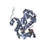
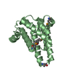
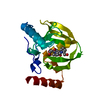
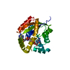
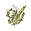
Entry Database : PDB / ID : 4umlTitle Crystal structure of ganglioside induced differentiation associated protein 2 (GDAP2) macro domain GANGLIOSIDE-INDUCED DIFFERENTIATION-ASSOCIATED PROTEIN 2 Keywords / Function / homology Function Domain/homology Component
/ / / / / / / / / / / / / / / / / / / / / / Biological species HOMO SAPIENS (human)Method / / / Resolution : 1.9 Å Authors Elkins, J.M. / Wang, J. / Kopec, J. / Wang, D. / Strain-Damerell, C. / Shrestha, L. / Sieg, C. / Tallant, C. / Newman, J.A. / von Delft, F. ...Elkins, J.M. / Wang, J. / Kopec, J. / Wang, D. / Strain-Damerell, C. / Shrestha, L. / Sieg, C. / Tallant, C. / Newman, J.A. / von Delft, F. / Bountra, C. / Edwards, A. / Knapp, S. Journal : To be Published Title : Structure of Gdap2Authors : Elkins, J.M. / Wang, J. / Knapp, S. History Deposition May 19, 2014 Deposition site / Processing site Revision 1.0 May 28, 2014 Provider / Type Revision 1.1 Jan 10, 2024 Group Data collection / Database references ... Data collection / Database references / Other / Refinement description Category chem_comp_atom / chem_comp_bond ... chem_comp_atom / chem_comp_bond / database_2 / pdbx_database_status / pdbx_initial_refinement_model Item / _database_2.pdbx_database_accession / _pdbx_database_status.status_code_sf
Show all Show less
 Movie
Movie Controller

 Yorodumi
 Open data
 Basic information
 Components
 Keywords
 Function and homology information
 HOMO SAPIENS (human)
 X-RAY DIFFRACTION /
  Authors
 Citation
 Structure visualization
 Downloads & links
 Other downloads
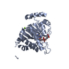


 PDBj
PDBj Assembly
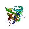

 SPODOPTERA FRUGIPERDA (fall armyworm) / References: UniProt: Q9NXN4
 Mass: 18.015 Da / Num. of mol.: 114 / Source method: isolated from a natural source / Formula: H2O
 Sample preparation
 / Beamline: I03 / Wavelength: 0.97625
  Processing