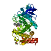
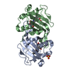
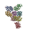
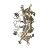
| Deposited unit | A: Putative acetyltransferase
B: Putative acetyltransferase
C: Putative acetyltransferase
D: Putative acetyltransferase
E: Putative acetyltransferase
F: Putative acetyltransferase
G: Putative acetyltransferase
H: Putative acetyltransferase
I: Putative acetyltransferase
hetero molecules
| Theoretical mass | Number of molelcules |
|---|
| Total (without water) | 144,457 | 20 |
|---|
| Polymers | 143,444 | 9 |
|---|
| Non-polymers | 1,013 | 11 |
|---|
| Water | 10,611 | 589 |
|---|
|
|---|
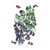
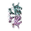
| 1 | A: Putative acetyltransferase
B: Putative acetyltransferase
hetero molecules
| Theoretical mass | Number of molelcules |
|---|
| Total (without water) | 32,061 | 4 |
|---|
| Polymers | 31,876 | 2 |
|---|
| Non-polymers | 184 | 2 |
|---|
| Water | 36 | 2 |
|---|
| Type | Name | Symmetry operation | Number |
|---|
| identity operation | 1_555 | x,y,z | 1 |
| Buried area | 5470 Å2 |
|---|
| ΔGint | -11 kcal/mol |
|---|
| Surface area | 12840 Å2 |
|---|
| Method | PISA |
|---|
|
|---|
| 2 | C: Putative acetyltransferase
D: Putative acetyltransferase
hetero molecules
| Theoretical mass | Number of molelcules |
|---|
| Total (without water) | 32,153 | 5 |
|---|
| Polymers | 31,876 | 2 |
|---|
| Non-polymers | 276 | 3 |
|---|
| Water | 36 | 2 |
|---|
| Type | Name | Symmetry operation | Number |
|---|
| identity operation | 1_555 | x,y,z | 1 |
| Buried area | 5900 Å2 |
|---|
| ΔGint | -15 kcal/mol |
|---|
| Surface area | 12810 Å2 |
|---|
| Method | PISA |
|---|
|
|---|
| 3 | E: Putative acetyltransferase
F: Putative acetyltransferase
hetero molecules
| Theoretical mass | Number of molelcules |
|---|
| Total (without water) | 32,153 | 5 |
|---|
| Polymers | 31,876 | 2 |
|---|
| Non-polymers | 276 | 3 |
|---|
| Water | 36 | 2 |
|---|
| Type | Name | Symmetry operation | Number |
|---|
| identity operation | 1_555 | x,y,z | 1 |
| Buried area | 5420 Å2 |
|---|
| ΔGint | -12 kcal/mol |
|---|
| Surface area | 12860 Å2 |
|---|
| Method | PISA |
|---|
|
|---|
| 4 | G: Putative acetyltransferase
H: Putative acetyltransferase
hetero molecules
| Theoretical mass | Number of molelcules |
|---|
| Total (without water) | 32,061 | 4 |
|---|
| Polymers | 31,876 | 2 |
|---|
| Non-polymers | 184 | 2 |
|---|
| Water | 36 | 2 |
|---|
| Type | Name | Symmetry operation | Number |
|---|
| identity operation | 1_555 | x,y,z | 1 |
| Buried area | 5420 Å2 |
|---|
| ΔGint | -13 kcal/mol |
|---|
| Surface area | 12830 Å2 |
|---|
| Method | PISA |
|---|
|
|---|
| 5 | I: Putative acetyltransferase
hetero molecules
I: Putative acetyltransferase
hetero molecules
| Theoretical mass | Number of molelcules |
|---|
| Total (without water) | 32,061 | 4 |
|---|
| Polymers | 31,876 | 2 |
|---|
| Non-polymers | 184 | 2 |
|---|
| Water | 36 | 2 |
|---|
| Type | Name | Symmetry operation | Number |
|---|
| identity operation | 1_555 | x,y,z | 1 | | crystal symmetry operation | 7_646 | y+1,x-1,-z+1 | 1 |
| Buried area | 5500 Å2 |
|---|
| ΔGint | -11 kcal/mol |
|---|
| Surface area | 12910 Å2 |
|---|
| Method | PISA |
|---|
|
|---|
| Unit cell | | Length a, b, c (Å) | 137.367, 137.367, 200.854 |
|---|
| Angle α, β, γ (deg.) | 90.00, 90.00, 90.00 |
|---|
| Int Tables number | 92 |
|---|
| Space group name H-M | P41212 |
|---|
|
|---|
 Movie
Movie Controller

 Yorodumi
 Open data
 Basic information
 Components
 Keywords
 Function and homology information
 Pseudomonas aeruginosa 18A (bacteria)
 X-RAY DIFFRACTION /
  Authors
 Citation
 Structure visualization
 Downloads & links
 Other downloads


 PDBj
PDBj

 Assembly
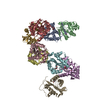


 Mass: 92.094 Da / Num. of mol.: 11 / Source method: obtained synthetically / Formula: C3H8O3
 Mass: 18.015 Da / Num. of mol.: 589 / Source method: isolated from a natural source / Formula: H2O
 Sample preparation
 / Beamline: 5C (4A) / Wavelength: 1 Å
 Processing