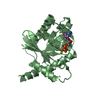
Entry Database : PDB / ID : 4uaiTitle Crystal structure of CXCL12 in complex with inhibitor Stromal cell-derived factor 1 Keywords / / / / / Function / homology Function Domain/homology Component
/ / / / / / / / / / / / / / / / / / / / / / / / / / / / / / / / / / / / / / / / / / / / / / / / / / / / / / / / / / / / / / / / / / / / / / / / / / Biological species Homo sapiens (human)Method / / Resolution : 1.9 Å Authors Smith, E.W. / Chen, Y. Funding support Organization Grant number Country National Institutes of Health/National Institute of General Medical Sciences (NIH/NIGMS) GM097381 National Institutes of Health/National Institute Of Allergy and Infectious Diseases (NIH/NIAID) AI058072 National Institutes of Health/National Cancer Institute (NIH/NCI) CA173056
Journal : J.Med.Chem. / Year : 2014Title : Structural Analysis of a Novel Small Molecule Ligand Bound to the CXCL12 Chemokine.Authors : Smith, E.W. / Liu, Y. / Getschman, A.E. / Peterson, F.C. / Ziarek, J.J. / Li, R. / Volkman, B.F. / Chen, Y. History Deposition Aug 9, 2014 Deposition site / Processing site Revision 1.0 Nov 12, 2014 Provider / Type Revision 1.1 Dec 3, 2014 Group Revision 2.0 Sep 27, 2017 Group Atomic model / Author supporting evidence ... Atomic model / Author supporting evidence / Database references / Derived calculations / Other / Refinement description / Source and taxonomy / Structure summary Category atom_site_anisotrop / citation ... atom_site_anisotrop / citation / entity_src_gen / pdbx_audit_support / pdbx_database_status / pdbx_struct_assembly / pdbx_struct_assembly_gen / pdbx_struct_assembly_prop / pdbx_struct_oper_list / software / struct_keywords Item _atom_site_anisotrop.pdbx_PDB_model_num / _atom_site_anisotrop.pdbx_label_asym_id ... _atom_site_anisotrop.pdbx_PDB_model_num / _atom_site_anisotrop.pdbx_label_asym_id / _atom_site_anisotrop.pdbx_label_atom_id / _atom_site_anisotrop.pdbx_label_comp_id / _atom_site_anisotrop.pdbx_label_seq_id / _citation.journal_id_CSD / _entity_src_gen.pdbx_alt_source_flag / _pdbx_audit_support.funding_organization / _pdbx_database_status.pdb_format_compatible / _pdbx_struct_assembly.oligomeric_details / _pdbx_struct_assembly_gen.asym_id_list / _pdbx_struct_assembly_prop.type / _pdbx_struct_assembly_prop.value / _pdbx_struct_oper_list.symmetry_operation / _software.classification / _struct_keywords.text Revision 2.1 Dec 4, 2019 Group / Category / Item Revision 2.2 Sep 27, 2023 Group / Database references / Refinement descriptionCategory chem_comp_atom / chem_comp_bond ... chem_comp_atom / chem_comp_bond / database_2 / pdbx_initial_refinement_model / refine_hist / struct_ncs_dom_lim Item _database_2.pdbx_DOI / _database_2.pdbx_database_accession ... _database_2.pdbx_DOI / _database_2.pdbx_database_accession / _refine_hist.number_atoms_solvent / _refine_hist.pdbx_number_atoms_ligand / _refine_hist.pdbx_number_atoms_nucleic_acid / _refine_hist.pdbx_number_atoms_protein / _struct_ncs_dom_lim.beg_auth_comp_id / _struct_ncs_dom_lim.beg_label_asym_id / _struct_ncs_dom_lim.beg_label_comp_id / _struct_ncs_dom_lim.beg_label_seq_id / _struct_ncs_dom_lim.end_auth_comp_id / _struct_ncs_dom_lim.end_label_asym_id / _struct_ncs_dom_lim.end_label_comp_id / _struct_ncs_dom_lim.end_label_seq_id Revision 2.3 Nov 13, 2024 Group / Category / pdbx_modification_feature
Show all Show less
 Movie
Movie Controller


 Open data
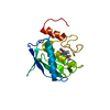
 Basic information
 Components
 Keywords
 Function and homology information
 Homo sapiens (human)
 X-RAY DIFFRACTION /
  Authors
 United States, 3items
  Citation
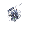
 Structure visualization
 Downloads & links
 Other downloads
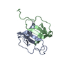


 PDBj
PDBj


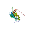
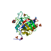
 Assembly

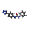
 Mass: 280.285 Da / Num. of mol.: 1 / Source method: obtained synthetically / Formula: C14H12N6O
 Mass: 96.063 Da / Num. of mol.: 2 / Source method: obtained synthetically / Formula: SO4
 Mass: 18.015 Da / Num. of mol.: 40 / Source method: isolated from a natural source / Formula: H2O
 Sample preparation
 Processing