Monochromator: Si(111) crystal / Protocol: SINGLE WAVELENGTH / Monochromatic (M) / Laue (L): M / Scattering type: x-ray
Radiation wavelength
Wavelength: 0.9788 Å / Relative weight: 1
Reflection
Resolution: 1.85→50 Å / Num. obs: 34839 / % possible obs: 99.4 % / Redundancy: 17.4 % / Biso Wilson estimate: 27.82 Å2 / Rmerge(I) obs: 0.067 / Net I/σ(I): 12.2
Reflection shell
Resolution: 1.85→1.92 Å / Redundancy: 16.7 % / Rmerge(I) obs: 0.595 / % possible all: 98.7
-
Processing
Software
Name
Version
Classification
PHENIX
refinement
HKL-3000
datacollection
HKL-3000
datascaling
PDB_EXTRACT
3.14
dataextraction
DENZO
datareduction
SCALEPACK
datascaling
Refinement
Method to determine structure: SAD / Resolution: 1.85→35.36 Å / SU ML: 0.24 / Cross valid method: THROUGHOUT / σ(F): 1.35 / Phase error: 31.33 / Stereochemistry target values: ML
Rfactor
Num. reflection
% reflection
Rfree
0.239
3352
4.97 %
Rwork
0.179
-
-
obs
0.182
67462
99.2 %
Solvent computation
Shrinkage radii: 0.9 Å / VDW probe radii: 1.11 Å / Solvent model: FLAT BULK SOLVENT MODEL
Displacement parameters
Biso mean: 65.44 Å2
Refinement step
Cycle: LAST / Resolution: 1.85→35.36 Å
Protein
Nucleic acid
Ligand
Solvent
Total
Num. atoms
3132
0
97
234
3463
Refine LS restraints
Refine-ID
Type
Dev ideal
Number
X-RAY DIFFRACTION
f_bond_d
0.007
3310
X-RAY DIFFRACTION
f_angle_d
1.078
4480
X-RAY DIFFRACTION
f_dihedral_angle_d
14.186
1181
X-RAY DIFFRACTION
f_chiral_restr
0.047
472
X-RAY DIFFRACTION
f_plane_restr
0.005
548
Refine LS restraints NCS
Ens-ID
Dom-ID
Auth asym-ID
Number
Refine-ID
Type
1
1
A
1615
X-RAY DIFFRACTION
POSITIONAL
1
2
B
1615
X-RAY DIFFRACTION
POSITIONAL
LS refinement shell
Resolution (Å)
Rfactor Rfree
Num. reflection Rfree
Rfactor Rwork
Num. reflection Rwork
Refine-ID
% reflection obs (%)
1.85-1.8784
0.3939
124
0.2729
2438
X-RAY DIFFRACTION
94
1.8784-1.9064
0.3813
137
0.2546
2686
X-RAY DIFFRACTION
98
1.9064-1.9362
0.2947
139
0.2438
2650
X-RAY DIFFRACTION
99
1.9362-1.9679
0.3157
141
0.2121
2677
X-RAY DIFFRACTION
99
1.9679-2.0019
0.3351
136
0.2072
2652
X-RAY DIFFRACTION
99
2.0019-2.0383
0.313
150
0.2053
2768
X-RAY DIFFRACTION
99
2.0383-2.0775
0.2261
130
0.1941
2607
X-RAY DIFFRACTION
99
2.0775-2.1199
0.2569
142
0.1887
2706
X-RAY DIFFRACTION
99
2.1199-2.166
0.3318
138
0.1947
2662
X-RAY DIFFRACTION
99
2.166-2.2163
0.2444
145
0.1933
2646
X-RAY DIFFRACTION
100
2.2163-2.2717
0.3863
140
0.1947
2750
X-RAY DIFFRACTION
99
2.2717-2.3332
0.3086
143
0.1937
2626
X-RAY DIFFRACTION
99
2.3332-2.4018
0.2884
136
0.1854
2672
X-RAY DIFFRACTION
100
2.4018-2.4793
0.2576
146
0.1805
2702
X-RAY DIFFRACTION
100
2.4793-2.5679
0.2655
141
0.197
2675
X-RAY DIFFRACTION
99
2.5679-2.6707
0.2802
142
0.2028
2741
X-RAY DIFFRACTION
100
2.6707-2.7922
0.3084
137
0.202
2668
X-RAY DIFFRACTION
100
2.7922-2.9393
0.3137
141
0.1969
2697
X-RAY DIFFRACTION
100
2.9393-3.1234
0.2354
139
0.1923
2648
X-RAY DIFFRACTION
100
3.1234-3.3643
0.2627
143
0.1869
2708
X-RAY DIFFRACTION
100
3.3643-3.7026
0.2097
139
0.1619
2676
X-RAY DIFFRACTION
100
3.7026-4.2376
0.1777
143
0.1606
2722
X-RAY DIFFRACTION
100
4.2376-5.336
0.1911
142
0.1355
2696
X-RAY DIFFRACTION
100
5.336-35.3655
0.1597
138
0.1681
2637
X-RAY DIFFRACTION
98
+
About Yorodumi
-
News
-
Feb 9, 2022. New format data for meta-information of EMDB entries
New format data for meta-information of EMDB entries
Version 3 of the EMDB header file is now the official format.
The previous official version 1.9 will be removed from the archive.
In the structure databanks used in Yorodumi, some data are registered as the other names, "COVID-19 virus" and "2019-nCoV". Here are the details of the virus and the list of structure data.
Jan 31, 2019. EMDB accession codes are about to change! (news from PDBe EMDB page)
EMDB accession codes are about to change! (news from PDBe EMDB page)
The allocation of 4 digits for EMDB accession codes will soon come to an end. Whilst these codes will remain in use, new EMDB accession codes will include an additional digit and will expand incrementally as the available range of codes is exhausted. The current 4-digit format prefixed with “EMD-” (i.e. EMD-XXXX) will advance to a 5-digit format (i.e. EMD-XXXXX), and so on. It is currently estimated that the 4-digit codes will be depleted around Spring 2019, at which point the 5-digit format will come into force.
The EM Navigator/Yorodumi systems omit the EMD- prefix.
Related info.:Q: What is EMD? / ID/Accession-code notation in Yorodumi/EM Navigator
Yorodumi is a browser for structure data from EMDB, PDB, SASBDB, etc.
This page is also the successor to EM Navigator detail page, and also detail information page/front-end page for Omokage search.
The word "yorodu" (or yorozu) is an old Japanese word meaning "ten thousand". "mi" (miru) is to see.
Related info.:EMDB / PDB / SASBDB / Comparison of 3 databanks / Yorodumi Search / Aug 31, 2016. New EM Navigator & Yorodumi / Yorodumi Papers / Jmol/JSmol / Function and homology information / Changes in new EM Navigator and Yorodumi
 Movie
Movie Controller


 Open data
 Basic information
 Components
 Keywords
 Function and homology information
 X-RAY DIFFRACTION /
  Authors
 United States, 1items
  Citation
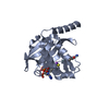
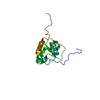
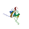
 Structure visualization
 Downloads & links
 Other downloads
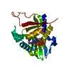


 PDBj
PDBj


 Assembly
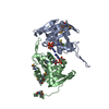

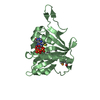


 Mass: 767.534 Da / Num. of mol.: 2 / Source method: obtained synthetically / Formula: C21H36N7O16P3S
 Mass: 35.453 Da / Num. of mol.: 1 / Source method: obtained synthetically / Formula: Cl
 Mass: 18.015 Da / Num. of mol.: 234 / Source method: isolated from a natural source / Formula: H2O
 Sample preparation
 Processing