 Movie
Movie Controller

[English] 日本語
 Yorodumi
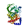
- PDB-4u4b: Crystal Structure of Pectate Lyase Pel3 from Pectobacterium carot... -
+ Open data

- Basic information
| Entry | Database: PDB / ID: 4u4b | ||||||
|---|---|---|---|---|---|---|---|
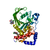
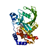
| Title | Crystal Structure of Pectate Lyase Pel3 from Pectobacterium carotovorum with one monomer in the A.U. | ||||||
 Components Components | Pectate lyase | ||||||
 Keywords Keywords | LYASE / Protein secretion / bacterial pathogenesis | ||||||
| Function / homology |  Function and homology information Function and homology informationpectate lyase / pectate lyase activity / pectin catabolic process / extracellular region / metal ion binding Similarity search - Function | ||||||
| Biological species |  Pectobacterium carotovorum (bacteria) Pectobacterium carotovorum (bacteria) | ||||||
| Method |  X-RAY DIFFRACTION / SYNCHROTRON / MOLECULAR REPLACEMENT / Resolution: 2.1 Å X-RAY DIFFRACTION / SYNCHROTRON / MOLECULAR REPLACEMENT / Resolution: 2.1 Å | ||||||
 Authors Authors | Ballut, L. / Gouet, P. / Shevchik, V.E. | ||||||
 Citation Citation | Journal: To Be Published Title: Crystal Structure of Pectate Lyase Pel3 from Pectobacterium carotovorum with one monomer in the A.U. Authors: Ballut, L. / Gouet, P. / Schevchik, V.E. / Pineau, C. / Guschinskaya, N. | ||||||
| History |
|
- Structure visualization
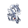
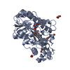
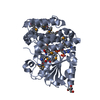
| Structure viewer | Molecule: MolmilJmol/JSmol |
|---|
- Downloads & links
-Download
| PDBx/mmCIF format | 4u4b.cif.gz | 79.3 KB | Display | PDBx/mmCIF format |
|---|---|---|---|---|
| PDB format | pdb4u4b.ent.gz | 57.3 KB | Display | PDB format |
| PDBx/mmJSON format | 4u4b.json.gz | Tree view | PDBx/mmJSON format | |
| Others |  Other downloads Other downloads |
-Validation report
| Arichive directory | https://data.pdbj.org/pub/pdb/validation_reports/u4/4u4bftp://data.pdbj.org/pub/pdb/validation_reports/u4/4u4b | HTTPS FTP |
|---|
-Related structure data
| Related structure data |  3b4nS S: Starting model for refinement |
|---|---|
| Similar structure data |


-Links
 PDBj
PDBj- Assembly
| Deposited unit | 
| ||||||||
|---|---|---|---|---|---|---|---|---|---|
| 1 |
| ||||||||
| Unit cell |
|
-Components
| #1: Protein | Mass: 37208.688 Da / Num. of mol.: 1 Source method: isolated from a genetically manipulated source Source: (gene. exp.) Pectobacterium carotovorum (bacteria) / Gene: pel-3 / Production host: |
|---|---|
| #2: Chemical | ChemComp-SO4 /   Mass: 96.063 Da / Num. of mol.: 1 / Source method: obtained synthetically / Formula: SO4 Mass: 96.063 Da / Num. of mol.: 1 / Source method: obtained synthetically / Formula: SO4 |
| #3: Chemical | ChemComp-EDO /   Mass: 62.068 Da / Num. of mol.: 1 / Source method: obtained synthetically / Formula: C2H6O2 Mass: 62.068 Da / Num. of mol.: 1 / Source method: obtained synthetically / Formula: C2H6O2 |
| #4: Water | ChemComp-HOH /  Mass: 18.015 Da / Num. of mol.: 183 / Source method: isolated from a natural source / Formula: H2O Mass: 18.015 Da / Num. of mol.: 183 / Source method: isolated from a natural source / Formula: H2O |
| Has protein modification | Y |
-Experimental details
-Experiment
| Experiment | Method: X-RAY DIFFRACTION |
|---|
- Sample preparation
| Crystal | Density Matthews: 1.92 Å3/Da / Density % sol: 35.4 % |
|---|---|
| Crystal grow | Temperature: 293 K / Method: vapor diffusion, sitting drop / pH: 7 Details: 1M succinic acid, 0.1M HEPES, 1% (m/v) PEG MME 2000 |
-Data collection
| Diffraction | Mean temperature: 100 K |
|---|---|
| Diffraction source | Source: SYNCHROTRON / Site: ESRF  / Beamline: ID29 / Wavelength: 0.939 Å / Beamline: ID29 / Wavelength: 0.939 Å |
| Detector | Type: DECTRIS PILATUS 6M / Detector: PIXEL / Date: Jul 2, 2013 |
| Radiation | Protocol: SINGLE WAVELENGTH / Monochromatic (M) / Laue (L): M / Scattering type: x-ray |
| Radiation wavelength | Wavelength: 0.939 Å / Relative weight: 1 |
| Reflection | Resolution: 2.1→19.7 Å / Num. all: 15124 / Num. obs: 15124 / % possible obs: 91.5 % / Observed criterion σ(F): 0 / Observed criterion σ(I): -3 / Redundancy: 2.87 % / Rmerge(I) obs: 0.054 / Rsym value: 0.054 / Net I/σ(I): 19.17 |
| Reflection shell | Resolution: 2.1→9.39 Å / Redundancy: 2.14 % / Rmerge(I) obs: 0.126 / Mean I/σ(I) obs: 6.71 / % possible all: 60.3 |
- Processing
| Software |
| ||||||||||||||||||||||||||||||||||||||||||
|---|---|---|---|---|---|---|---|---|---|---|---|---|---|---|---|---|---|---|---|---|---|---|---|---|---|---|---|---|---|---|---|---|---|---|---|---|---|---|---|---|---|---|---|
| Refinement | Method to determine structure: MOLECULAR REPLACEMENT Starting model: 3B4N Resolution: 2.1→19.7 Å / SU ML: 0.26 / Cross valid method: FREE R-VALUE / σ(F): 1.36 / Phase error: 27.15 / Stereochemistry target values: ML
| ||||||||||||||||||||||||||||||||||||||||||
| Solvent computation | Shrinkage radii: 0.9 Å / VDW probe radii: 1.11 Å / Solvent model: FLAT BULK SOLVENT MODEL | ||||||||||||||||||||||||||||||||||||||||||
| Refinement step | Cycle: LAST / Resolution: 2.1→19.7 Å
| ||||||||||||||||||||||||||||||||||||||||||
| Refine LS restraints |
| ||||||||||||||||||||||||||||||||||||||||||
| LS refinement shell |
|
