 Movie
Movie Controller

[English] 日本語
 Yorodumi
- PDB-4tw1: Crystal structure of the octameric pore complex of the Staphyloco... -
+ Open data

- Basic information
| Entry | Database: PDB / ID: 4tw1 | ||||||
|---|---|---|---|---|---|---|---|
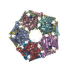
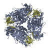
| Title | Crystal structure of the octameric pore complex of the Staphylococcus aureus Bi-component Toxin LukGH | ||||||
 Components Components | (Possible leukocidin subunit) x 2 | ||||||
 Keywords Keywords | TOXIN / octamer leukocidin pore-forming toxin | ||||||
| Function / homology | Leukocidin/porin MspA / Leukocidin-like / Distorted Sandwich / Mainly Beta / : / :  Function and homology information Function and homology information | ||||||
| Biological species |   Staphylococcus aureus (bacteria) Staphylococcus aureus (bacteria) | ||||||
| Method |  X-RAY DIFFRACTION / SYNCHROTRON / MOLECULAR REPLACEMENT / molecular replacement / Resolution: 2.8 Å X-RAY DIFFRACTION / SYNCHROTRON / MOLECULAR REPLACEMENT / molecular replacement / Resolution: 2.8 Å | ||||||
 Authors Authors | Logan, D.T. / Hakansson, M. / Saline, M. / Kimbung, R. / Badarau, A. / Rouha, H. / Nagy, E. | ||||||
 Citation Citation | Journal: J.Biol.Chem. / Year: 2015 Title: Structure-Function Analysis of Heterodimer Formation, Oligomerization, and Receptor Binding of the Staphylococcus aureus Bi-component Toxin LukGH. Authors: Badarau, A. / Rouha, H. / Malafa, S. / Logan, D.T. / Hakansson, M. / Stulik, L. / Dolezilkova, I. / Teubenbacher, A. / Gross, K. / Maierhofer, B. / Weber, S. / Jagerhofer, M. / Hoffman, D. / Nagy, E. | ||||||
| History |
|
- Structure visualization
| Structure viewer | Molecule: MolmilJmol/JSmol |
|---|
- Downloads & links
-Download
| PDBx/mmCIF format | 4tw1.cif.gz | 910.6 KB | Display | PDBx/mmCIF format |
|---|---|---|---|---|
| PDB format | pdb4tw1.ent.gz | 753.2 KB | Display | PDB format |
| PDBx/mmJSON format | 4tw1.json.gz | Tree view | PDBx/mmJSON format | |
| Others |  Other downloads Other downloads |
-Validation report
| Arichive directory | https://data.pdbj.org/pub/pdb/validation_reports/tw/4tw1ftp://data.pdbj.org/pub/pdb/validation_reports/tw/4tw1 | HTTPS FTP |
|---|
-Related structure data
| Related structure data |  3b07S S: Starting model for refinement |
|---|---|
| Similar structure data |

-Links
 PDBj
PDBj- Assembly
| Deposited unit | 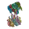
| ||||||||
|---|---|---|---|---|---|---|---|---|---|
| 1 | 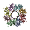
| ||||||||
| 2 | 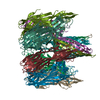
| ||||||||
| Unit cell |
|
-Components
| #1: Protein | Mass: 35825.539 Da / Num. of mol.: 8 Source method: isolated from a genetically manipulated source Source: (gene. exp.) Staphylococcus aureus (bacteria) / Strain: USA300 / TCH1516 / Gene: USA300HOU_2011 / Production host: #2: Protein | Mass: 37682.680 Da / Num. of mol.: 8 / Fragment: Residues 28-351 Source method: isolated from a genetically manipulated source Source: (gene. exp.) Staphylococcus aureus (bacteria) / Strain: USA300 / TCH1516 / Gene: USA300HOU_2013 / Production host: #3: Water | ChemComp-HOH / |  Mass: 18.015 Da / Num. of mol.: 205 / Source method: isolated from a natural source / Formula: H2O Mass: 18.015 Da / Num. of mol.: 205 / Source method: isolated from a natural source / Formula: H2O |
|---|
-Experimental details
-Experiment
| Experiment | Method: X-RAY DIFFRACTION / Number of used crystals: 1 |
|---|
- Sample preparation
| Crystal | Density Matthews: 4.39 Å3/Da / Density % sol: 71.96 % / Description: Thin plates |
|---|---|
| Crystal grow | Temperature: 293 K / Method: vapor diffusion, sitting drop / pH: 7.5 Details: Drops consisted of 100 nl protein in 20 mM HEPES buffer pH 7.5 and 100 nl reservoir 30 mM MgCl2, 30 mM CaCl2, 0.1 M Na HEPES/MOPS buffer pH 7.5, 12.5 % v/v MPD, 12.5 % w/v PEG 1000, 12.5 % ...Details: Drops consisted of 100 nl protein in 20 mM HEPES buffer pH 7.5 and 100 nl reservoir 30 mM MgCl2, 30 mM CaCl2, 0.1 M Na HEPES/MOPS buffer pH 7.5, 12.5 % v/v MPD, 12.5 % w/v PEG 1000, 12.5 % w/v PEG 3350. Crystals appeared after 7 days. |
-Data collection
| Diffraction | Mean temperature: 100 K | ||||||||||||||||||||||||||||||||||||||||||||||||||||||||||||||||||||||||||||||||||||||||||||||||||||||||||||||
|---|---|---|---|---|---|---|---|---|---|---|---|---|---|---|---|---|---|---|---|---|---|---|---|---|---|---|---|---|---|---|---|---|---|---|---|---|---|---|---|---|---|---|---|---|---|---|---|---|---|---|---|---|---|---|---|---|---|---|---|---|---|---|---|---|---|---|---|---|---|---|---|---|---|---|---|---|---|---|---|---|---|---|---|---|---|---|---|---|---|---|---|---|---|---|---|---|---|---|---|---|---|---|---|---|---|---|---|---|---|---|---|
| Diffraction source | Source: SYNCHROTRON / Site: MAX II  / Beamline: I911-3 / Wavelength: 1 Å / Beamline: I911-3 / Wavelength: 1 Å | ||||||||||||||||||||||||||||||||||||||||||||||||||||||||||||||||||||||||||||||||||||||||||||||||||||||||||||||
| Detector | Type: MARMOSAIC 225 mm CCD / Detector: CCD / Date: Dec 12, 2013 | ||||||||||||||||||||||||||||||||||||||||||||||||||||||||||||||||||||||||||||||||||||||||||||||||||||||||||||||
| Radiation | Monochromator: Double crystal / Protocol: SINGLE WAVELENGTH / Monochromatic (M) / Laue (L): M / Scattering type: x-ray | ||||||||||||||||||||||||||||||||||||||||||||||||||||||||||||||||||||||||||||||||||||||||||||||||||||||||||||||
| Radiation wavelength | Wavelength: 1 Å / Relative weight: 1 | ||||||||||||||||||||||||||||||||||||||||||||||||||||||||||||||||||||||||||||||||||||||||||||||||||||||||||||||
| Reflection | Resolution: 2.8→48.188 Å / Num. all: 222018 / Num. obs: 222018 / % possible obs: 97.9 % / Redundancy: 3.1 % / Biso Wilson estimate: 56.4 Å2 / Rpim(I) all: 0.109 / Rrim(I) all: 0.194 / Rsym value: 0.16 / Net I/av σ(I): 4.824 / Net I/σ(I): 10.1 / Num. measured all: 685808 | ||||||||||||||||||||||||||||||||||||||||||||||||||||||||||||||||||||||||||||||||||||||||||||||||||||||||||||||
| Reflection shell | Diffraction-ID: 1 / Rejects: _
|
-Phasing
| Phasing | Method: molecular replacement | |||||||||
|---|---|---|---|---|---|---|---|---|---|---|
| Phasing MR | Model details: Phaser MODE: MR_AUTO
|
- Processing
| Software |
| ||||||||||||||||||||||||||||||||||||||||||||||||||||||||||||
|---|---|---|---|---|---|---|---|---|---|---|---|---|---|---|---|---|---|---|---|---|---|---|---|---|---|---|---|---|---|---|---|---|---|---|---|---|---|---|---|---|---|---|---|---|---|---|---|---|---|---|---|---|---|---|---|---|---|---|---|---|---|
| Refinement | Method to determine structure: MOLECULAR REPLACEMENT Starting model: 3B07 prepared using CHAINSAW Resolution: 2.8→48 Å / Cor.coef. Fo:Fc: 0.906 / Cor.coef. Fo:Fc free: 0.856 / WRfactor Rfree: 0.2318 / WRfactor Rwork: 0.1798 / FOM work R set: 0.6812 / SU B: 20.998 / SU ML: 0.362 / SU R Cruickshank DPI: 0.512 / SU Rfree: 0.3468 / Cross valid method: THROUGHOUT / σ(F): 0 / ESU R: 0.512 / ESU R Free: 0.347 / Stereochemistry target values: MAXIMUM LIKELIHOOD / Details: U VALUES : REFINED INDIVIDUALLY
| ||||||||||||||||||||||||||||||||||||||||||||||||||||||||||||
| Solvent computation | Ion probe radii: 0.8 Å / Shrinkage radii: 0.8 Å / VDW probe radii: 1.2 Å / Solvent model: MASK | ||||||||||||||||||||||||||||||||||||||||||||||||||||||||||||
| Displacement parameters | Biso max: 132.12 Å2 / Biso mean: 36.001 Å2 / Biso min: 2 Å2
| ||||||||||||||||||||||||||||||||||||||||||||||||||||||||||||
| Refinement step | Cycle: final / Resolution: 2.8→48 Å
| ||||||||||||||||||||||||||||||||||||||||||||||||||||||||||||
| Refine LS restraints |
| ||||||||||||||||||||||||||||||||||||||||||||||||||||||||||||
| LS refinement shell | Resolution: 2.8→2.869 Å
|
