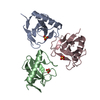
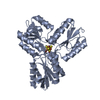
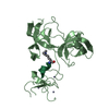
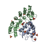
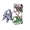
E3ubiquitin-proteinligaseUHRF2 / Np95/ICBP90-like RING finger protein / Np95-like RING finger protein / Nuclear protein 97 / Nuclear ...Np95/ICBP90-like RING finger protein / Np95-like RING finger protein / Nuclear protein 97 / Nuclear zinc finger protein Np97 / RING finger protein 107 / Ubiquitin-like PHD and RING finger domain-containing protein 2 / Ubiquitin-like-containing PHD and RING finger domains protein 2
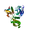
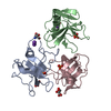
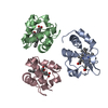
Mass: 31359.346 Da / Num. of mol.: 1 Fragment: Tandem Tudor and PHD domains, UNP residues 109-395 Mutation: deletion from 169 to 176, E383A Source method: isolated from a genetically manipulated source Source: (gene. exp.) Homo sapiens (human) / Gene: UHRF2, NIRF, RNF107 / Plasmid: pET28-MHL / Production host: Escherichia coli (E. coli) / Strain (production host): BL21(DE3)-V2R-pRARE2 References: UniProt: Q96PU4, Ligases; Forming carbon-nitrogen bonds; Acid-amino-acid ligases (peptide synthases)
Resolution: 2.29→50 Å / Cor.coef. Fo:Fc: 0.943 / Cor.coef. Fo:Fc free: 0.927 / SU B: 13.81 / SU ML: 0.177 / Cross valid method: THROUGHOUT / ESU R: 0.287 / ESU R Free: 0.222 / Stereochemistry target values: MAXIMUM LIKELIHOOD / Details: HYDROGENS HAVE BEEN ADDED IN THE RIDING POSITIONS
Rfactor
Num. reflection
% reflection
Selection details
Rfree
0.25982
721
5.1 %
RANDOM
Rwork
0.22902
-
-
-
obs
0.23067
13552
98.53 %
-
Solvent computation
Ion probe radii: 0.8 Å / Shrinkage radii: 0.8 Å / VDW probe radii: 1.2 Å / Solvent model: MASK
 Movie
Movie Controller


 Open data
 Basic information
 Components
 Keywords
 Function and homology information
 Homo sapiens (human)
 X-RAY DIFFRACTION /
  Authors
 Citation
 Structure visualization
 Downloads & links
 Other downloads
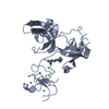


 PDBj
PDBj


 Assembly
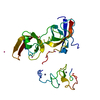


 Mass: 65.409 Da / Num. of mol.: 3 / Source method: obtained synthetically / Formula: Zn
 Num. of mol.: 7 / Source method: obtained synthetically
 Mass: 18.015 Da / Num. of mol.: 53 / Source method: isolated from a natural source / Formula: H2O
 Sample preparation
 / Beamline: 19-ID / Wavelength: 1.2822 Å
 Processing