Mass: 18.015 Da / Num. of mol.: 36 / Source method: isolated from a natural source / Formula: H2O
-
Experimental details
-
Experiment
Experiment
Method: X-RAY DIFFRACTION
-
Sample preparation
Crystal
Density Matthews: 2.03 Å3/Da / Density % sol: 39.37 %
Crystal grow
Temperature: 293.15 K / Method: vapor diffusion, hanging drop / pH: 8.5 Details: Citrate synthase crystals grew at 20 degrees Celsius in 0.2M Magnesium chloride hexahydrate, 0.1M Tris pH=8.5, 3.4M 1,6-Hexandiol in a 75/25 protein/reservoir-ratio drop of 1 microlitre and ...Details: Citrate synthase crystals grew at 20 degrees Celsius in 0.2M Magnesium chloride hexahydrate, 0.1M Tris pH=8.5, 3.4M 1,6-Hexandiol in a 75/25 protein/reservoir-ratio drop of 1 microlitre and with a protein concentration of 5.2 mg/ml.
Resolution: 2.6→47.14 Å / Cor.coef. Fo:Fc: 0.937 / Cor.coef. Fo:Fc free: 0.932 / SU B: 4.225 / SU ML: 0.102 / Cross valid method: THROUGHOUT / ESU R: 0.051 / ESU R Free: 0.04 / Stereochemistry target values: MAXIMUM LIKELIHOOD / Details: HYDROGENS HAVE BEEN ADDED IN THE RIDING POSITIONS
Rfactor
Num. reflection
% reflection
Selection details
Rfree
0.19405
1290
5.4 %
RANDOM
Rwork
0.17378
-
-
-
obs
0.17494
22819
99.04 %
-
Solvent computation
Ion probe radii: 0.8 Å / Shrinkage radii: 0.8 Å / VDW probe radii: 1.2 Å / Solvent model: MASK
 Movie
Movie Controller


 Open data
 Basic information
 Components
 Keywords
 Function and homology information
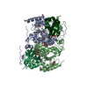
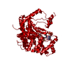
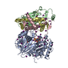
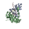
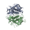
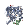
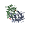
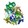
 Mycobacterium tuberculosis (bacteria)
 X-RAY DIFFRACTION /
  Authors
 Italy, 1items
  Citation
 Structure visualization
 Downloads & links
 Other downloads

 PDBj
PDBj


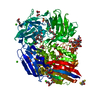
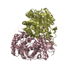
 Assembly
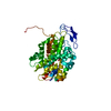


 Mass: 131.064 Da / Num. of mol.: 1 / Source method: obtained synthetically / Formula: C4H3O5
 Mass: 18.015 Da / Num. of mol.: 36 / Source method: isolated from a natural source / Formula: H2O
 Sample preparation
 / Beamline: ID29 / Wavelength: 0.9725 Å
 Processing