| Entry | Database: PDB / ID: 4tv7
|
|---|
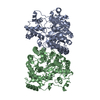
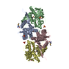
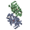
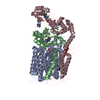
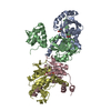
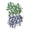
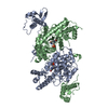
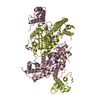
| Title | Crystal structure of Bacillus subtilis GabR at 2.05 Angstroms resolution |
|---|
 Components Components | HTH-type transcriptional regulatory protein GabR |
|---|
 Keywords Keywords | TRANSCRIPTION / transcriptional regulator |
|---|
| Function / homology |  Function and homology information Function and homology information
: / Transcription regulator HTH, GntR / Bacterial regulatory proteins, gntR family / GntR-type HTH domain profile. / helix_turn_helix gluconate operon transcriptional repressor / Aminotransferase, class I/classII / Aminotransferase class I and II / Aspartate Aminotransferase; domain 2 / Type I PLP-dependent aspartate aminotransferase-like (Major domain) / Winged helix-like DNA-binding domain superfamily/Winged helix DNA-binding domain ...: / Transcription regulator HTH, GntR / Bacterial regulatory proteins, gntR family / GntR-type HTH domain profile. / helix_turn_helix gluconate operon transcriptional repressor / Aminotransferase, class I/classII / Aminotransferase class I and II / Aspartate Aminotransferase; domain 2 / Type I PLP-dependent aspartate aminotransferase-like (Major domain) / Winged helix-like DNA-binding domain superfamily/Winged helix DNA-binding domain / Pyridoxal phosphate-dependent transferase, major domain / Pyridoxal phosphate-dependent transferase / Arc Repressor Mutant, subunit A / Winged helix DNA-binding domain superfamily / Winged helix-like DNA-binding domain superfamily / Orthogonal Bundle / 3-Layer(aba) Sandwich / Mainly Alpha / Alpha BetaSimilarity search - Domain/homology |
|---|
| Biological species |   Bacillus subtilis 168 (bacteria) Bacillus subtilis 168 (bacteria) |
|---|
| Method |  X-RAY DIFFRACTION / SYNCHROTRON / MOLECULAR REPLACEMENT / MIR / Resolution: 2.05 Å X-RAY DIFFRACTION / SYNCHROTRON / MOLECULAR REPLACEMENT / MIR / Resolution: 2.05 Å |
|---|
 Authors Authors | Goto, M. / Okuda, K. / Yoshimura, T. |
|---|
 Citation Citation | Journal: Mol. Microbiol. / Year: 2015
Title: Role of the aminotransferase domain in Bacillus subtilis GabR, a pyridoxal 5'-phosphate-dependent transcriptional regulator
Authors: Okuda, K. / Kato, S. / Ito, T. / Shiraki, S. / Kawase, Y. / Goto, M. / Kawashima, S. / Hemmi, H. / Fukada, H. / Yoshimura, T. |
|---|
| History | | Deposition | Jun 26, 2014 | Deposition site: RCSB / Processing site: PDBJ |
|---|
| Revision 1.0 | Dec 24, 2014 | Provider: repository / Type: Initial release |
|---|
| Revision 1.1 | Mar 15, 2017 | Group: Database references |
|---|
| Revision 1.2 | Jan 29, 2020 | Group: Data collection / Category: diffrn_source / Item: _diffrn_source.pdbx_synchrotron_site |
|---|
| Revision 1.3 | Apr 9, 2025 | Group: Data collection / Database references / Structure summary
Category: chem_comp_atom / chem_comp_bond ...chem_comp_atom / chem_comp_bond / database_2 / pdbx_entry_details / pdbx_modification_feature
Item: _database_2.pdbx_DOI / _database_2.pdbx_database_accession |
|---|
|
|---|
 Movie
Movie Controller

 Yorodumi
 Open data
 Basic information
 Structure visualization
 Downloads & links
 Other downloads
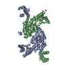


 PDBj
PDBj Assembly
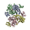
 Mass: 18.015 Da / Num. of mol.: 563 / Source method: isolated from a natural source / Formula: H2O
 Sample preparation
 / Beamline: BL-5A / Wavelength: 1 Å
 Processing