 Movie
Movie Controller

[English] 日本語
 Yorodumi
- PDB-4tqk: Structural basis of specific recognition of non-reducing terminal... -
+ Open data

- Basic information
| Entry | Database: PDB / ID: 4tqk | ||||||
|---|---|---|---|---|---|---|---|
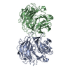
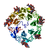
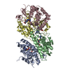
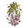
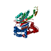
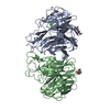
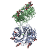
| Title | Structural basis of specific recognition of non-reducing terminal N-acetylglucosamine by an Agrocybe aegerita lection | ||||||
 Components Components | Lectin 2 | ||||||
 Keywords Keywords | SUGAR BINDING PROTEIN / complex / lectin / GlcNAc | ||||||
| Function / homology | FG-GAP-like repeat / FG-GAP repeat / Integrin alpha, N-terminal / Lectin 2 Function and homology information Function and homology information | ||||||
| Biological species |  Agrocybe aegerita (fungus) Agrocybe aegerita (fungus) | ||||||
| Method |  X-RAY DIFFRACTION / SYNCHROTRON / Resolution: 2.1 Å X-RAY DIFFRACTION / SYNCHROTRON / Resolution: 2.1 Å | ||||||
 Authors Authors | Hu, Y.L. / Ren, X.M. / Li, D.F. / Jiang, S. / Lan, X.Q. / Sun, H. / Wang, D.C. | ||||||
 Citation Citation | Journal: Plos One / Year: 2015 Title: Structural Basis of Specific Recognition of Non-Reducing Terminal N-Acetylglucosamine by an Agrocybe aegerita Lectin. Authors: Ren, X.M. / Li, D.F. / Jiang, S. / Lan, X.Q. / Hu, Y. / Sun, H. / Wang, D.C. | ||||||
| History |
|
- Structure visualization
| Structure viewer | Molecule: MolmilJmol/JSmol |
|---|
- Downloads & links
-Download
| PDBx/mmCIF format | 4tqk.cif.gz | 185.4 KB | Display | PDBx/mmCIF format |
|---|---|---|---|---|
| PDB format | pdb4tqk.ent.gz | 144.8 KB | Display | PDB format |
| PDBx/mmJSON format | 4tqk.json.gz | Tree view | PDBx/mmJSON format | |
| Others |  Other downloads Other downloads |
-Validation report
| Arichive directory | https://data.pdbj.org/pub/pdb/validation_reports/tq/4tqkftp://data.pdbj.org/pub/pdb/validation_reports/tq/4tqk | HTTPS FTP |
|---|
-Related structure data


-Links
 PDBj
PDBj
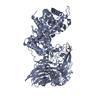
- Assembly
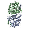
| Deposited unit | 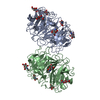
| ||||||||
|---|---|---|---|---|---|---|---|---|---|
| 1 |
| ||||||||
| Unit cell |
|
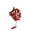
-Components
| #1: Protein | Mass: 43082.289 Da / Num. of mol.: 2 / Mutation: I126T Source method: isolated from a genetically manipulated source Source: (gene. exp.) Agrocybe aegerita (fungus) / Gene: aal-2 / Plasmid: pET30a / Production host:  #2: Sugar | ChemComp-NAG /   Type: D-saccharide, beta linking / Mass: 221.208 Da / Num. of mol.: 11 / Source method: obtained synthetically / Formula: C8H15NO6 Type: D-saccharide, beta linking / Mass: 221.208 Da / Num. of mol.: 11 / Source method: obtained synthetically / Formula: C8H15NO6#3: Water | ChemComp-HOH / |  Mass: 18.015 Da / Num. of mol.: 874 / Source method: isolated from a natural source / Formula: H2O Mass: 18.015 Da / Num. of mol.: 874 / Source method: isolated from a natural source / Formula: H2O |
|---|
-Experimental details
-Experiment
| Experiment | Method: X-RAY DIFFRACTION / Number of used crystals: 1 |
|---|
- Sample preparation
| Crystal | Density Matthews: 2.3 Å3/Da / Density % sol: 46.62 % |
|---|---|
| Crystal grow | Temperature: 298 K / Method: vapor diffusion, hanging drop / pH: 6 Details: 25% PEG 4000 (w/v), 6% Tacsimate pH 6.0 and 0.1M MES pH 6.0 |
-Data collection
| Diffraction | Mean temperature: 100 K | ||||||||||||||||||||||||||||||||||||||||||||||||||||||||||||||||||||||||||||||||||||||||||||||||||||||||||||||
|---|---|---|---|---|---|---|---|---|---|---|---|---|---|---|---|---|---|---|---|---|---|---|---|---|---|---|---|---|---|---|---|---|---|---|---|---|---|---|---|---|---|---|---|---|---|---|---|---|---|---|---|---|---|---|---|---|---|---|---|---|---|---|---|---|---|---|---|---|---|---|---|---|---|---|---|---|---|---|---|---|---|---|---|---|---|---|---|---|---|---|---|---|---|---|---|---|---|---|---|---|---|---|---|---|---|---|---|---|---|---|---|
| Diffraction source | Source: SYNCHROTRON / Site: BSRF  / Beamline: 1W2B / Wavelength: 1 Å / Beamline: 1W2B / Wavelength: 1 Å | ||||||||||||||||||||||||||||||||||||||||||||||||||||||||||||||||||||||||||||||||||||||||||||||||||||||||||||||
| Detector | Type: RIGAKU RAXIS / Detector: IMAGE PLATE / Date: Jan 1, 2012 | ||||||||||||||||||||||||||||||||||||||||||||||||||||||||||||||||||||||||||||||||||||||||||||||||||||||||||||||
| Radiation | Protocol: SINGLE WAVELENGTH / Monochromatic (M) / Laue (L): M / Scattering type: x-ray | ||||||||||||||||||||||||||||||||||||||||||||||||||||||||||||||||||||||||||||||||||||||||||||||||||||||||||||||
| Radiation wavelength | Wavelength: 1 Å / Relative weight: 1 | ||||||||||||||||||||||||||||||||||||||||||||||||||||||||||||||||||||||||||||||||||||||||||||||||||||||||||||||
| Reflection | Resolution: 2.1→85.754 Å / Num. all: 41489 / Num. obs: 41489 / % possible obs: 88.8 % / Redundancy: 3 % / Rpim(I) all: 0.085 / Rrim(I) all: 0.166 / Rsym value: 0.141 / Net I/av σ(I): 5.185 / Net I/σ(I): 6.3 / Num. measured all: 126025 | ||||||||||||||||||||||||||||||||||||||||||||||||||||||||||||||||||||||||||||||||||||||||||||||||||||||||||||||
| Reflection shell | Diffraction-ID: 1 / Rejects: _
|
- Processing
| Software |
| |||||||||||||||||||||||||||||||||||||||||||||||||||||||||||||||||||||||||||||||||||||||||||||||||||||||||
|---|---|---|---|---|---|---|---|---|---|---|---|---|---|---|---|---|---|---|---|---|---|---|---|---|---|---|---|---|---|---|---|---|---|---|---|---|---|---|---|---|---|---|---|---|---|---|---|---|---|---|---|---|---|---|---|---|---|---|---|---|---|---|---|---|---|---|---|---|---|---|---|---|---|---|---|---|---|---|---|---|---|---|---|---|---|---|---|---|---|---|---|---|---|---|---|---|---|---|---|---|---|---|---|---|---|---|
| Refinement | Resolution: 2.1→26.28 Å / FOM work R set: 0.8568 / SU ML: 0.29 / Cross valid method: FREE R-VALUE / σ(F): 0 / Phase error: 21.47 / Stereochemistry target values: ML
| |||||||||||||||||||||||||||||||||||||||||||||||||||||||||||||||||||||||||||||||||||||||||||||||||||||||||
| Solvent computation | Shrinkage radii: 0.83 Å / VDW probe radii: 1.1 Å / Solvent model: FLAT BULK SOLVENT MODEL / Bsol: 26.619 Å2 / ksol: 0.371 e/Å3 | |||||||||||||||||||||||||||||||||||||||||||||||||||||||||||||||||||||||||||||||||||||||||||||||||||||||||
| Displacement parameters | Biso max: 61.18 Å2 / Biso mean: 12.39 Å2 / Biso min: 2.17 Å2
| |||||||||||||||||||||||||||||||||||||||||||||||||||||||||||||||||||||||||||||||||||||||||||||||||||||||||
| Refinement step | Cycle: final / Resolution: 2.1→26.28 Å
| |||||||||||||||||||||||||||||||||||||||||||||||||||||||||||||||||||||||||||||||||||||||||||||||||||||||||
| Refine LS restraints |
| |||||||||||||||||||||||||||||||||||||||||||||||||||||||||||||||||||||||||||||||||||||||||||||||||||||||||
| LS refinement shell | Refine-ID: X-RAY DIFFRACTION / Total num. of bins used: 14
|
