 Movie
Movie Controller

[English] 日本語
 Yorodumi
- PDB-4tq7: N-terminal domain of C. Reinhardtii SAS-6 homolog bld12p Q93E (NN10) -
+ Open data

- Basic information
| Entry | Database: PDB / ID: 4tq7 | ||||||
|---|---|---|---|---|---|---|---|
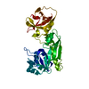
| Title | N-terminal domain of C. Reinhardtii SAS-6 homolog bld12p Q93E (NN10) | ||||||
 Components Components | Centriole protein | ||||||
 Keywords Keywords | STRUCTURAL PROTEIN / centriole SAS-6 / cartwheel / beta-sandwich / alpha-beta protein / centriolar | ||||||
| Function / homology | : / Sas-6-like, oligomerization domain / Spindle assembly abnormal protein 6, N-terminal / SAS-6, N-terminal domain superfamily / Centriolar protein SAS N-terminal domain / identical protein binding / Centriole protein Function and homology information Function and homology information | ||||||
| Biological species |   Chlamydomonas reinhardtii (plant) Chlamydomonas reinhardtii (plant) | ||||||
| Method |  X-RAY DIFFRACTION / SYNCHROTRON / MOLECULAR REPLACEMENT / Resolution: 2.643 Å X-RAY DIFFRACTION / SYNCHROTRON / MOLECULAR REPLACEMENT / Resolution: 2.643 Å | ||||||
 Authors Authors | Hilbert, M. / Kraatz, S.H.W. | ||||||
| Funding support |  Switzerland, 1items Switzerland, 1items
| ||||||
 Citation Citation | Journal: Nat.Cell Biol. / Year: 2016 Title: SAS-6 engineering reveals interdependence between cartwheel and microtubules in determining centriole architecture. Authors: Hilbert, M. / Noga, A. / Frey, D. / Hamel, V. / Guichard, P. / Kraatz, S.H. / Pfreundschuh, M. / Hosner, S. / Fluckiger, I. / Jaussi, R. / Wieser, M.M. / Thieltges, K.M. / Deupi, X. / ...Authors: Hilbert, M. / Noga, A. / Frey, D. / Hamel, V. / Guichard, P. / Kraatz, S.H. / Pfreundschuh, M. / Hosner, S. / Fluckiger, I. / Jaussi, R. / Wieser, M.M. / Thieltges, K.M. / Deupi, X. / Muller, D.J. / Kammerer, R.A. / Gonczy, P. / Hirono, M. / Steinmetz, M.O. #1: Journal: Cell / Year: 2011Title: Structural basis of the 9-fold symmetry of centrioles. Authors: Kitagawa, D. / Vakonakis, I. / Olieric, N. / Hilbert, M. / Keller, D. / Olieric, V. / Bortfeld, M. / Erat, M.C. / Flueckiger, I. / Goenczy, P. / Steinmetz, M.O. | ||||||
| History |
|
- Structure visualization
| Structure viewer | Molecule: MolmilJmol/JSmol |
|---|
- Downloads & links
-Download
| PDBx/mmCIF format | 4tq7.cif.gz | 72.4 KB | Display | PDBx/mmCIF format |
|---|---|---|---|---|
| PDB format | pdb4tq7.ent.gz | 52.5 KB | Display | PDB format |
| PDBx/mmJSON format | 4tq7.json.gz | Tree view | PDBx/mmJSON format | |
| Others |  Other downloads Other downloads |
-Validation report
| Arichive directory | https://data.pdbj.org/pub/pdb/validation_reports/tq/4tq7ftp://data.pdbj.org/pub/pdb/validation_reports/tq/4tq7 | HTTPS FTP |
|---|
-Related structure data
| Related structure data |  4to7C  4tpzC  4ttwC  4ttxC  4ttyC  4ttzC  4u2iC  4u2jC  3q0yS C: citing same article ( S: Starting model for refinement |
|---|---|
| Similar structure data |


-Links
 PDBj
PDBj- Assembly
| Deposited unit | 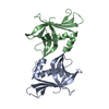
| ||||||||
|---|---|---|---|---|---|---|---|---|---|
| 1 |
| ||||||||
| Unit cell |
|
-Components
| #1: Protein | Mass: 18104.605 Da / Num. of mol.: 2 / Mutation: Q93E Source method: isolated from a genetically manipulated source Source: (gene. exp.) Chlamydomonas reinhardtii (plant) / Gene: CrSAS-6 / Production host:  #2: Water | ChemComp-HOH / |  Mass: 18.015 Da / Num. of mol.: 84 / Source method: isolated from a natural source / Formula: H2O Mass: 18.015 Da / Num. of mol.: 84 / Source method: isolated from a natural source / Formula: H2O |
|---|
-Experimental details
-Experiment
| Experiment | Method: X-RAY DIFFRACTION |
|---|
- Sample preparation
| Crystal | Density Matthews: 2.22 Å3/Da / Density % sol: 44.68 % |
|---|---|
| Crystal grow | Temperature: 293 K / Method: evaporation / pH: 6.5 / Details: PEG3350, MES-NaOH |
-Data collection
| Diffraction | Mean temperature: 100 K |
|---|---|
| Diffraction source | Source: SYNCHROTRON / Site: SLS / Beamline: X06DA / Wavelength: 1 Å |
| Detector | Type: DECTRIS PILATUS 2M-F / Detector: PIXEL / Date: Aug 8, 2012 |
| Radiation | Protocol: SINGLE WAVELENGTH / Monochromatic (M) / Laue (L): M / Scattering type: x-ray |
| Radiation wavelength | Wavelength: 1 Å / Relative weight: 1 |
| Reflection | Resolution: 2.643→41.435 Å / Num. obs: 9912 / % possible obs: 99.7 % / Redundancy: 12.5 % / Net I/σ(I): 25.06 |
| Reflection shell | Resolution: 2.64→2.71 Å / Redundancy: 11.9 % / Rmerge(I) obs: 0.537 / Mean I/σ(I) obs: 6.26 / % possible all: 97.1 |
- Processing
| Software |
| |||||||||||||||||||||||||||||||||||
|---|---|---|---|---|---|---|---|---|---|---|---|---|---|---|---|---|---|---|---|---|---|---|---|---|---|---|---|---|---|---|---|---|---|---|---|---|
| Refinement | Method to determine structure: MOLECULAR REPLACEMENT Starting model: 3q0y Resolution: 2.643→41.435 Å / SU ML: 0.28 / Cross valid method: FREE R-VALUE / σ(F): 1.99 / Phase error: 27.5 / Stereochemistry target values: ML
| |||||||||||||||||||||||||||||||||||
| Solvent computation | Shrinkage radii: 0.9 Å / VDW probe radii: 1.11 Å / Solvent model: FLAT BULK SOLVENT MODEL | |||||||||||||||||||||||||||||||||||
| Refinement step | Cycle: LAST / Resolution: 2.643→41.435 Å
| |||||||||||||||||||||||||||||||||||
| Refine LS restraints |
| |||||||||||||||||||||||||||||||||||
| LS refinement shell |
|
