| Entry | Database: PDB / ID: 4tll
|
|---|
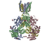
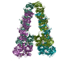
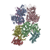
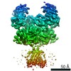
| Title | Crystal structure of GluN1/GluN2B NMDA receptor, structure 1 |
|---|
 Components Components | (receptor subunit ...) x 2 |
|---|
 Keywords Keywords | SIGNALING PROTEIN / Neurotransmitter receptor / NMDA receptor / GluN1/GluN2B / Membrane protein / Ion channel |
|---|
| Function / homology |  Function and homology information Function and homology information
NMDA glutamate receptor activity / NMDA selective glutamate receptor complex / response to zinc ion / ligand-gated sodium channel activity / response to magnesium ion / ligand-gated monoatomic ion channel activity / glutamate-gated calcium ion channel activity / ionotropic glutamate receptor signaling pathway / sodium ion transmembrane transport / synaptic transmission, glutamatergic ...NMDA glutamate receptor activity / NMDA selective glutamate receptor complex / response to zinc ion / ligand-gated sodium channel activity / response to magnesium ion / ligand-gated monoatomic ion channel activity / glutamate-gated calcium ion channel activity / ionotropic glutamate receptor signaling pathway / sodium ion transmembrane transport / synaptic transmission, glutamatergic / transmitter-gated monoatomic ion channel activity involved in regulation of postsynaptic membrane potential / regulation of membrane potential / excitatory postsynaptic potential / regulation of synaptic plasticity / postsynaptic density membrane / calcium ion transmembrane transport / long-term synaptic potentiation / late endosome / signaling receptor activity / chemical synaptic transmission / postsynaptic membrane / lysosome / neuron projection / synapse / metal ion binding / plasma membraneSimilarity search - Function Glutamate [NMDA] receptor, epsilon subunit, C-terminal / N-methyl D-aspartate receptor 2B3 C-terminus / : / : / Response regulator / Ionotropic glutamate receptor, metazoa / Ligated ion channel L-glutamate- and glycine-binding site / Ligand-gated ion channel / Ionotropic glutamate receptor, L-glutamate and glycine-binding domain / Ligated ion channel L-glutamate- and glycine-binding site ...Glutamate [NMDA] receptor, epsilon subunit, C-terminal / N-methyl D-aspartate receptor 2B3 C-terminus / : / : / Response regulator / Ionotropic glutamate receptor, metazoa / Ligated ion channel L-glutamate- and glycine-binding site / Ligand-gated ion channel / Ionotropic glutamate receptor, L-glutamate and glycine-binding domain / Ligated ion channel L-glutamate- and glycine-binding site / : / Ionotropic glutamate receptor / Eukaryotic homologues of bacterial periplasmic substrate binding proteins. / Periplasmic binding protein-like II / Receptor, ligand binding region / Receptor family ligand binding region / D-Maltodextrin-Binding Protein; domain 2 / Periplasmic binding protein-like I / Rossmann fold / 3-Layer(aba) Sandwich / Alpha BetaSimilarity search - Domain/homology 1-AMINOCYCLOPROPANECARBOXYLIC ACID / trans-1-aminocyclobutane-1,3-dicarboxylic acid / Chem-QEM / Glutamate receptor ionotropic, NMDA 1 / Glutamate receptor ionotropic, NMDA 2B / Glutamate receptor ionotropic, NMDA 1Similarity search - Component |
|---|
| Biological species |  Xenopus laevis (African clawed frog) Xenopus laevis (African clawed frog) |
|---|
| Method |  X-RAY DIFFRACTION / SYNCHROTRON / MOLECULAR REPLACEMENT / Resolution: 3.59 Å X-RAY DIFFRACTION / SYNCHROTRON / MOLECULAR REPLACEMENT / Resolution: 3.59 Å |
|---|
 Authors Authors | Gouaux, E. / Lee, C.-H. / Lu, W. |
|---|
 Citation Citation | Journal: Nature / Year: 2014
Title: NMDA receptor structures reveal subunit arrangement and pore architecture.
Authors: Lee, C.H. / Lu, W. / Michel, J.C. / Goehring, A. / Du, J. / Song, X. / Gouaux, E. |
|---|
| History | | Deposition | May 30, 2014 | Deposition site: RCSB / Processing site: RCSB |
|---|
| Revision 1.0 | Jul 2, 2014 | Provider: repository / Type: Initial release |
|---|
| Revision 1.1 | Jul 9, 2014 | Group: Database references / Structure summary |
|---|
| Revision 1.2 | Jul 16, 2014 | Group: Database references |
|---|
| Revision 1.3 | Sep 24, 2014 | Group: Database references |
|---|
| Revision 1.4 | Jul 29, 2020 | Group: Data collection / Database references ...Data collection / Database references / Derived calculations / Other / Refinement description / Source and taxonomy / Structure summary
Category: chem_comp / citation ...chem_comp / citation / entity / entity_src_gen / pdbx_chem_comp_identifier / pdbx_database_status / pdbx_entity_nonpoly / pdbx_struct_oper_list / refine_hist / struct_conn / struct_site / struct_site_gen
Item: _chem_comp.name / _chem_comp.type ..._chem_comp.name / _chem_comp.type / _citation.journal_id_CSD / _entity.pdbx_description / _entity_src_gen.pdbx_alt_source_flag / _pdbx_database_status.pdb_format_compatible / _pdbx_entity_nonpoly.name / _pdbx_struct_oper_list.symmetry_operation / _struct_conn.pdbx_role
Description: Carbohydrate remediation / Provider: repository / Type: Remediation |
|---|
| Revision 1.5 | Dec 27, 2023 | Group: Data collection / Database references / Structure summary
Category: chem_comp / chem_comp_atom ...chem_comp / chem_comp_atom / chem_comp_bond / database_2
Item: _chem_comp.pdbx_synonyms / _database_2.pdbx_DOI / _database_2.pdbx_database_accession |
|---|
|
|---|
 Movie
Movie Controller


 Open data
 Basic information
 Structure visualization
 Downloads & links
 Other downloads
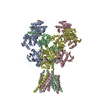


 PDBj
PDBj

 Assembly
 Homo sapiens (human) / References: UniProt: C0KD18, UniProt: A0A1L8F5J9*PLUS
 Type: D-saccharide, beta linking / Mass: 221.208 Da / Num. of mol.: 4 / Source method: obtained synthetically / Formula: C8H15NO6


 Type: peptide linking / Mass: 101.104 Da / Num. of mol.: 1 / Source method: obtained synthetically / Formula: C4H7NO2
 Mass: 339.471 Da / Num. of mol.: 2 / Source method: obtained synthetically / Formula: C22H29NO2
 Mass: 159.140 Da / Num. of mol.: 1 / Source method: obtained synthetically / Formula: C6H9NO4
 Sample preparation
 / Beamline: 8.2.1 / Wavelength: 1 Å
 Processing