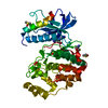
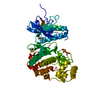
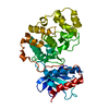
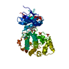
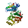
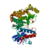
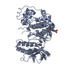
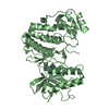
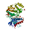
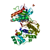
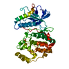
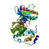
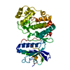
[English] 日本語
Yorodumi
- PDB-4s32: Crystal structure of ERK2 AMP-PNP complex -

+
Open data


ID or keywords:

Loading...

-
Basic information

Entry
Database: PDB / ID: 4s32
TitleCrystal structure of ERK2 AMP-PNP complex
ComponentsMitogen-activated protein kinase 1
KeywordsTRANSFERASE / signal transduction / phosphorylation
Function / homology
Function and homology information


phospho-PLA2 pathway / RAF-independent MAPK1/3 activation / MAPK1 (ERK2) activation / Signaling by NODAL / Frs2-mediated activation / ERK/MAPK targets / ERKs are inactivated / Activation of the AP-1 family of transcription factors / Transcriptional and post-translational regulation of MITF-M expression and activity / Negative feedback regulation of MAPK pathway ...phospho-PLA2 pathway / RAF-independent MAPK1/3 activation / MAPK1 (ERK2) activation / Signaling by NODAL / Frs2-mediated activation / ERK/MAPK targets / ERKs are inactivated / Activation of the AP-1 family of transcription factors / Transcriptional and post-translational regulation of MITF-M expression and activity / Negative feedback regulation of MAPK pathway / Gastrin-CREB signalling pathway via PKC and MAPK / Estrogen-dependent nuclear events downstream of ESR-membrane signaling / IFNG signaling activates MAPKs / Golgi Cisternae Pericentriolar Stack Reorganization / RHO GTPases Activate WASPs and WAVEs / Estrogen-stimulated signaling through PRKCZ / Growth hormone receptor signaling / Spry regulation of FGF signaling / SMAD2/SMAD3:SMAD4 heterotrimer regulates transcription / Oxidative Stress Induced Senescence / Senescence-Associated Secretory Phenotype (SASP) / Oncogene Induced Senescence / Regulation of actin dynamics for phagocytic cup formation / Downregulation of SMAD2/3:SMAD4 transcriptional activity / Signal attenuation / NCAM signaling for neurite out-growth / Negative regulation of FGFR1 signaling / Negative regulation of FGFR3 signaling / Negative regulation of FGFR4 signaling / Regulation of the apoptosome activity / Signaling by Activin / Negative regulation of FGFR2 signaling / Signal transduction by L1 / RHO GTPases Activate NADPH Oxidases / Negative regulation of MAPK pathway / PI5P, PP2A and IER3 Regulate PI3K/AKT Signaling / Interferon gamma signaling / FCERI mediated MAPK activation / Regulation of HSF1-mediated heat shock response / diadenosine tetraphosphate biosynthetic process / MAP2K and MAPK activation / neural crest cell development / Recycling pathway of L1 / cellular response to toxic substance / cardiac neural crest cell development involved in heart development / caveolin-mediated endocytosis / cytosine metabolic process / mitogen-activated protein kinase kinase kinase binding / cellular response to methionine / response to epidermal growth factor / positive regulation of macrophage proliferation / response to alcohol / regulation of cellular pH / outer ear morphogenesis / regulation of Golgi inheritance / labyrinthine layer blood vessel development / RAF/MAP kinase cascade / Thrombin signalling through proteinase activated receptors (PARs) / ERBB signaling pathway / Neutrophil degranulation / mammary gland epithelial cell proliferation / trachea formation / regulation of early endosome to late endosome transport / regulation of stress-activated MAPK cascade / cellular response to insulin-like growth factor stimulus / ERBB2-ERBB3 signaling pathway / positive regulation of macrophage chemotaxis / regulation of cytoskeleton organization / response to exogenous dsRNA / face development / lung morphogenesis / positive regulation of telomere maintenance / response to testosterone / pseudopodium / Bergmann glial cell differentiation / androgen receptor signaling pathway / thyroid gland development / steroid hormone receptor signaling pathway / decidualization / MAP kinase activity / negative regulation of cell differentiation / regulation of ossification / JUN kinase activity / mitogen-activated protein kinase / phosphatase binding / Schwann cell development / progesterone receptor signaling pathway / stress-activated MAPK cascade / estrous cycle / positive regulation of cardiac muscle cell proliferation / cellular response to platelet-derived growth factor stimulus / cellular response to cAMP / sensory perception of pain / dendrite cytoplasm / phosphotyrosine residue binding / myelination / RNA polymerase II CTD heptapeptide repeat kinase activity / peptidyl-threonine phosphorylation / ERK1 and ERK2 cascade / cellular response to epidermal growth factor stimulus
Similarity search - Function
Mitogen-activated protein (MAP) kinase, ERK1/2 / Mitogen-activated protein (MAP) kinase, conserved site / MAP kinase signature. / : / Phosphorylase Kinase; domain 1 / Phosphorylase Kinase; domain 1 / Transferase(Phosphotransferase) domain 1 / Transferase(Phosphotransferase); domain 1 / Serine/threonine-protein kinase, active site / Serine/Threonine protein kinases active-site signature. ...Mitogen-activated protein (MAP) kinase, ERK1/2 / Mitogen-activated protein (MAP) kinase, conserved site / MAP kinase signature. / : / Phosphorylase Kinase; domain 1 / Phosphorylase Kinase; domain 1 / Transferase(Phosphotransferase) domain 1 / Transferase(Phosphotransferase); domain 1 / Serine/threonine-protein kinase, active site / Serine/Threonine protein kinases active-site signature. / Protein kinase domain / Serine/Threonine protein kinases, catalytic domain / Protein kinase, ATP binding site / Protein kinases ATP-binding region signature. / Protein kinase domain profile. / Protein kinase domain / Protein kinase-like domain superfamily / 2-Layer Sandwich / Orthogonal Bundle / Mainly Alpha / Alpha Beta
Similarity search - Domain/homology
PHOSPHOAMINOPHOSPHONIC ACID-ADENYLATE ESTER / Mitogen-activated protein kinase 1
Similarity search - Component
Biological speciesRattus norvegicus (Norway rat)
MethodX-RAY DIFFRACTION / SYNCHROTRON / MOLECULAR REPLACEMENT / Resolution: 1.34 Å
AuthorsLivnah, O. / Karamansha, Y. / Tzarum, N.
CitationJournal: To be Published
Title: Crystal structure of ERK2 AMP-PNP complex
Authors: Livnah, O. / Karamansha, Y.
History
DepositionJan 26, 2015Deposition site: RCSB / Processing site: RCSB
Revision 1.0Jan 27, 2016Provider: repository / Type: Initial release
Revision 1.1Sep 20, 2023Group: Data collection / Database references ...Data collection / Database references / Derived calculations / Refinement description
Category: chem_comp_atom / chem_comp_bond ...chem_comp_atom / chem_comp_bond / database_2 / pdbx_initial_refinement_model / pdbx_struct_conn_angle / struct_conn / struct_site
Item: _database_2.pdbx_DOI / _database_2.pdbx_database_accession ..._database_2.pdbx_DOI / _database_2.pdbx_database_accession / _pdbx_struct_conn_angle.ptnr1_auth_comp_id / _pdbx_struct_conn_angle.ptnr1_auth_seq_id / _pdbx_struct_conn_angle.ptnr1_label_asym_id / _pdbx_struct_conn_angle.ptnr1_label_atom_id / _pdbx_struct_conn_angle.ptnr1_label_comp_id / _pdbx_struct_conn_angle.ptnr1_label_seq_id / _pdbx_struct_conn_angle.ptnr3_auth_comp_id / _pdbx_struct_conn_angle.ptnr3_auth_seq_id / _pdbx_struct_conn_angle.ptnr3_label_asym_id / _pdbx_struct_conn_angle.ptnr3_label_atom_id / _pdbx_struct_conn_angle.ptnr3_label_comp_id / _pdbx_struct_conn_angle.ptnr3_label_seq_id / _struct_conn.pdbx_dist_value / _struct_conn.ptnr1_auth_comp_id / _struct_conn.ptnr1_auth_seq_id / _struct_conn.ptnr1_label_asym_id / _struct_conn.ptnr1_label_atom_id / _struct_conn.ptnr1_label_comp_id / _struct_conn.ptnr1_label_seq_id / _struct_conn.ptnr2_auth_comp_id / _struct_conn.ptnr2_auth_seq_id / _struct_conn.ptnr2_label_asym_id / _struct_conn.ptnr2_label_atom_id / _struct_conn.ptnr2_label_comp_id / _struct_site.pdbx_auth_asym_id / _struct_site.pdbx_auth_comp_id / _struct_site.pdbx_auth_seq_id

-
Structure visualization

Structure viewerMolecule:
MolmilJmol/JSmol

Downloads & links

-
Assembly

Deposited unit
A: Mitogen-activated protein kinase 1
hetero molecules


Theoretical massNumber of molelcules
Total (without water)41,9574
Polymers41,3311
Non-polymers6273
Water3,819212
1


  • Idetical with deposited unit
  • defined by author&software
TypeNameSymmetry operationNumber
identity operation1_555x,y,z1
MethodPISA
Unit cell
Length a, b, c (Å)48.694, 69.968, 59.822
Angle α, β, γ (deg.)90.00, 108.98, 90.00
Int Tables number4
Space group name H-MP1211

-
Components

#1: Protein Mitogen-activated protein kinase 1 / MAP kinase 1 / MAPK 1 / ERT1 / Extracellular signal-regulated kinase 2 / ERK-2 / MAP kinase isoform ...MAP kinase 1 / MAPK 1 / ERT1 / Extracellular signal-regulated kinase 2 / ERK-2 / MAP kinase isoform p42 / p42-MAPK / Mitogen-activated protein kinase 2 / MAP kinase 2 / MAPK 2


Mass: 41330.555 Da / Num. of mol.: 1
Source method: isolated from a genetically manipulated source
Source: (gene. exp.) Rattus norvegicus (Norway rat) / Gene: Mapk1, Erk2, Mapk, Prkm1 / Production host: Escherichia coli (E. coli)
References: UniProt: P63086, mitogen-activated protein kinase
#2: Chemical ChemComp-ANP / PHOSPHOAMINOPHOSPHONIC ACID-ADENYLATE ESTER


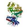
Mass: 506.196 Da / Num. of mol.: 1 / Source method: obtained synthetically / Formula: C10H17N6O12P3 / Comment: AMP-PNP, energy-carrying molecule analogue*YM
#3: Chemical ChemComp-SO4 / SULFATE ION


Mass: 96.063 Da / Num. of mol.: 1 / Source method: obtained synthetically / Formula: SO4
#4: Chemical ChemComp-MG / MAGNESIUM ION


Mass: 24.305 Da / Num. of mol.: 1 / Source method: obtained synthetically / Formula: Mg
#5: Water ChemComp-HOH / water


Mass: 18.015 Da / Num. of mol.: 212 / Source method: isolated from a natural source / Formula: H2O

-
Experimental details

-
Experiment

ExperimentMethod: X-RAY DIFFRACTION

-
Sample preparation

CrystalDensity Matthews: 2.33 Å3/Da / Density % sol: 47.25 %
Crystal growTemperature: 293 K / Method: vapor diffusion, sitting drop / pH: 7
Details: 61 mM Ammonium Sulfate, 100mM Bis Tris pH 7.0, 12-15% (v/v) PEG 3350, VAPOR DIFFUSION, SITTING DROP, temperature 293K

-
Data collection

DiffractionMean temperature: 100 K
Diffraction sourceSource: SYNCHROTRON / Site: ESRF / Beamline: ID14-4 / Wavelength: 0.94 Å
DetectorType: ADSC QUANTUM 315r / Detector: CCD / Date: Feb 20, 2010
RadiationMonochromator: optical hutch / Protocol: SINGLE WAVELENGTH / Monochromatic (M) / Laue (L): M / Scattering type: x-ray
Radiation wavelengthWavelength: 0.94 Å / Relative weight: 1
ReflectionResolution: 1.34→50 Å / Num. all: 83034 / Num. obs: 83034 / % possible obs: 98.3 % / Observed criterion σ(F): 0 / Observed criterion σ(I): 0

-
Processing

Software
NameVersionClassification
HKL-2000data collection
AMoREphasing
REFMAC5.7.0029refinement
HKL-2000data reduction
HKL-2000data scaling
RefinementMethod to determine structure: MOLECULAR REPLACEMENT
Starting model: 1ERK

1erk
PDB Unreleased entry


Resolution: 1.34→28.6 Å / Cor.coef. Fo:Fc: 0.968 / Cor.coef. Fo:Fc free: 0.955 / SU B: 1.191 / SU ML: 0.048 / Cross valid method: THROUGHOUT / ESU R: 0.058 / ESU R Free: 0.062 / Stereochemistry target values: MAXIMUM LIKELIHOOD / Details: HYDROGENS HAVE BEEN ADDED IN THE RIDING POSITIONS
RfactorNum. reflection% reflectionSelection details
Rfree0.2306 4135 5 %RANDOM
Rwork0.19787 ---
obs0.19948 78872 97.84 %-
all-78872 --
Solvent computationIon probe radii: 0.8 Å / Shrinkage radii: 0.8 Å / VDW probe radii: 1.2 Å / Solvent model: MASK
Displacement parametersBiso mean: 31.251 Å2
Baniso -1Baniso -2Baniso -3
1-0.21 Å20 Å20.27 Å2
2---0.38 Å20 Å2
3---0 Å2
Refinement stepCycle: LAST / Resolution: 1.34→28.6 Å
ProteinNucleic acidLigandSolventTotal
Num. atoms2807 0 29 212 3048
Refine LS restraints
Refine-IDTypeDev idealDev ideal targetNumber
X-RAY DIFFRACTIONr_bond_refined_d0.0260.0192904
X-RAY DIFFRACTIONr_bond_other_d0.0020.022782
X-RAY DIFFRACTIONr_angle_refined_deg2.3471.9833943
X-RAY DIFFRACTIONr_angle_other_deg0.9936410
X-RAY DIFFRACTIONr_dihedral_angle_1_deg6.0835344
X-RAY DIFFRACTIONr_dihedral_angle_2_deg41.78724.234137
X-RAY DIFFRACTIONr_dihedral_angle_3_deg14.75915507
X-RAY DIFFRACTIONr_dihedral_angle_4_deg21.6241516
X-RAY DIFFRACTIONr_chiral_restr0.1420.2436
X-RAY DIFFRACTIONr_gen_planes_refined0.0110.0213222
X-RAY DIFFRACTIONr_gen_planes_other0.0010.02663
X-RAY DIFFRACTIONr_nbd_refined
X-RAY DIFFRACTIONr_nbd_other
X-RAY DIFFRACTIONr_nbtor_refined
X-RAY DIFFRACTIONr_nbtor_other
X-RAY DIFFRACTIONr_xyhbond_nbd_refined
X-RAY DIFFRACTIONr_xyhbond_nbd_other
X-RAY DIFFRACTIONr_metal_ion_refined
X-RAY DIFFRACTIONr_metal_ion_other
X-RAY DIFFRACTIONr_symmetry_vdw_refined
X-RAY DIFFRACTIONr_symmetry_vdw_other
X-RAY DIFFRACTIONr_symmetry_hbond_refined
X-RAY DIFFRACTIONr_symmetry_hbond_other
X-RAY DIFFRACTIONr_symmetry_metal_ion_refined
X-RAY DIFFRACTIONr_symmetry_metal_ion_other
X-RAY DIFFRACTIONr_mcbond_it2.7222.8421382
X-RAY DIFFRACTIONr_mcbond_other2.7212.841381
X-RAY DIFFRACTIONr_mcangle_it3.6184.2481724
X-RAY DIFFRACTIONr_mcangle_other
X-RAY DIFFRACTIONr_scbond_it4.2073.2851522
X-RAY DIFFRACTIONr_scbond_other
X-RAY DIFFRACTIONr_scangle_it
X-RAY DIFFRACTIONr_scangle_other
X-RAY DIFFRACTIONr_long_range_B_refined
X-RAY DIFFRACTIONr_long_range_B_other
X-RAY DIFFRACTIONr_rigid_bond_restr
X-RAY DIFFRACTIONr_sphericity_free
X-RAY DIFFRACTIONr_sphericity_bonded
LS refinement shellResolution: 1.341→1.376 Å / Total num. of bins used: 20
RfactorNum. reflection% reflection
Rfree0.339 278 -
Rwork0.321 5387 -
obs--90.44 %

+
About Yorodumi

-
News

-
Feb 9, 2022. New format data for meta-information of EMDB entries

New format data for meta-information of EMDB entries

  • Version 3 of the EMDB header file is now the official format.
  • The previous official version 1.9 will be removed from the archive.

Related info.:EMDB header

External links:wwPDB to switch to version 3 of the EMDB data model

-
Aug 12, 2020. Covid-19 info

Covid-19 info

URL: https://pdbj.org/emnavi/covid19.php

New page: Covid-19 featured information page in EM Navigator.

Related info.:Covid-19 info / Mar 5, 2020. Novel coronavirus structure data

+
Mar 5, 2020. Novel coronavirus structure data

Novel coronavirus structure data

Related info.:Yorodumi Speices / Aug 12, 2020. Covid-19 info

External links:COVID-19 featured content - PDBj / Molecule of the Month (242):Coronavirus Proteases

+
Jan 31, 2019. EMDB accession codes are about to change! (news from PDBe EMDB page)

EMDB accession codes are about to change! (news from PDBe EMDB page)

  • The allocation of 4 digits for EMDB accession codes will soon come to an end. Whilst these codes will remain in use, new EMDB accession codes will include an additional digit and will expand incrementally as the available range of codes is exhausted. The current 4-digit format prefixed with “EMD-” (i.e. EMD-XXXX) will advance to a 5-digit format (i.e. EMD-XXXXX), and so on. It is currently estimated that the 4-digit codes will be depleted around Spring 2019, at which point the 5-digit format will come into force.
  • The EM Navigator/Yorodumi systems omit the EMD- prefix.

Related info.:Q: What is EMD? / ID/Accession-code notation in Yorodumi/EM Navigator

External links:EMDB Accession Codes are Changing Soon! / Contact to PDBj

+
Jul 12, 2017. Major update of PDB

Major update of PDB

  • wwPDB released updated PDB data conforming to the new PDBx/mmCIF dictionary.
  • This is a major update changing the version number from 4 to 5, and with Remediation, in which all the entries are updated.
  • In this update, many items about electron microscopy experimental information are reorganized (e.g. em_software).
  • Now, EM Navigator and Yorodumi are based on the updated data.

External links:wwPDB Remediation / Enriched Model Files Conforming to OneDep Data Standards Now Available in the PDB FTP Archive

-
Yorodumi

Thousand views of thousand structures

  • Yorodumi is a browser for structure data from EMDB, PDB, SASBDB, etc.
  • This page is also the successor to EM Navigator detail page, and also detail information page/front-end page for Omokage search.
  • The word "yorodu" (or yorozu) is an old Japanese word meaning "ten thousand". "mi" (miru) is to see.

Related info.:EMDB / PDB / SASBDB / Comparison of 3 databanks / Yorodumi Search / Aug 31, 2016. New EM Navigator & Yorodumi / Yorodumi Papers / Jmol/JSmol / Function and homology information / Changes in new EM Navigator and Yorodumi

Read more