 Movie
Movie Controller

[English] 日本語
 Yorodumi
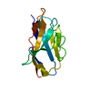
- PDB-4s1z: Crystal structure of TRABID NZF1 in complex with K29 linked di-Ub... -
+ Open data

- Basic information
| Entry | Database: PDB / ID: 4s1z | ||||||
|---|---|---|---|---|---|---|---|
| Title | Crystal structure of TRABID NZF1 in complex with K29 linked di-Ubiquitin | ||||||
 Components Components |
| ||||||
 Keywords Keywords | HYDROLASE / zinc finger / Protease / Ubiquitin binding | ||||||
| Function / homology |  Function and homology information Function and homology informationprotein K33-linked deubiquitination / protein K29-linked deubiquitination / protein deubiquitination involved in ubiquitin-dependent protein catabolic process / Ovarian tumor domain proteases / Degradation of beta-catenin by the destruction complex / regulation of cell morphogenesis / protein K63-linked deubiquitination / K63-linked polyubiquitin modification-dependent protein binding / Peptide chain elongation / Selenocysteine synthesis ...protein K33-linked deubiquitination / protein K29-linked deubiquitination / protein deubiquitination involved in ubiquitin-dependent protein catabolic process / Ovarian tumor domain proteases / Degradation of beta-catenin by the destruction complex / regulation of cell morphogenesis / protein K63-linked deubiquitination / K63-linked polyubiquitin modification-dependent protein binding / Peptide chain elongation / Selenocysteine synthesis / Formation of a pool of free 40S subunits / Eukaryotic Translation Termination / positive regulation of Wnt signaling pathway / Response of EIF2AK4 (GCN2) to amino acid deficiency / SRP-dependent cotranslational protein targeting to membrane / Viral mRNA Translation / Nonsense Mediated Decay (NMD) independent of the Exon Junction Complex (EJC) / GTP hydrolysis and joining of the 60S ribosomal subunit / L13a-mediated translational silencing of Ceruloplasmin expression / Major pathway of rRNA processing in the nucleolus and cytosol / Nonsense Mediated Decay (NMD) enhanced by the Exon Junction Complex (EJC) / Maturation of protein E / cytoskeleton organization / Maturation of protein E / ER Quality Control Compartment (ERQC) / Myoclonic epilepsy of Lafora / IRAK2 mediated activation of TAK1 complex / Alpha-protein kinase 1 signaling pathway / FLT3 signaling by CBL mutants / IRAK1 recruits IKK complex / IRAK1 recruits IKK complex upon TLR7/8 or 9 stimulation / Prevention of phagosomal-lysosomal fusion / Glycogen synthesis / cytosolic ribosome / IRAK2 mediated activation of TAK1 complex upon TLR7/8 or 9 stimulation / Endosomal Sorting Complex Required For Transport (ESCRT) / Regulation of TBK1, IKKε (IKBKE)-mediated activation of IRF3, IRF7 / TICAM1,TRAF6-dependent induction of TAK1 complex / Membrane binding and targetting of GAG proteins / Regulation of TBK1, IKKε-mediated activation of IRF3, IRF7 upon TLR3 ligation / Negative regulation of FLT3 / Constitutive Signaling by NOTCH1 HD Domain Mutants / PTK6 Regulates RTKs and Their Effectors AKT1 and DOK1 / Regulation of FZD by ubiquitination / TICAM1-dependent activation of IRF3/IRF7 / NOTCH2 Activation and Transmission of Signal to the Nucleus / p75NTR recruits signalling complexes / APC/C:Cdc20 mediated degradation of Cyclin B / VLDLR internalisation and degradation / Downregulation of ERBB4 signaling / TRAF6-mediated induction of TAK1 complex within TLR4 complex / TRAF6 mediated IRF7 activation in TLR7/8 or 9 signaling / APC-Cdc20 mediated degradation of Nek2A / Regulation of innate immune responses to cytosolic DNA / NF-kB is activated and signals survival / InlA-mediated entry of Listeria monocytogenes into host cells / Regulation of pyruvate metabolism / Downregulation of ERBB2:ERBB3 signaling / NRIF signals cell death from the nucleus / Pexophagy / Activated NOTCH1 Transmits Signal to the Nucleus / Regulation of PTEN localization / Regulation of BACH1 activity / Synthesis of active ubiquitin: roles of E1 and E2 enzymes / TICAM1, RIP1-mediated IKK complex recruitment / Translesion synthesis by REV1 / MAP3K8 (TPL2)-dependent MAPK1/3 activation / Translesion synthesis by POLK / Activation of IRF3, IRF7 mediated by TBK1, IKKε (IKBKE) / Downregulation of TGF-beta receptor signaling / Translesion synthesis by POLI / IKK complex recruitment mediated by RIP1 / Regulation of activated PAK-2p34 by proteasome mediated degradation / JNK (c-Jun kinases) phosphorylation and activation mediated by activated human TAK1 / Gap-filling DNA repair synthesis and ligation in GG-NER / Josephin domain DUBs / InlB-mediated entry of Listeria monocytogenes into host cell / PINK1-PRKN Mediated Mitophagy / TGF-beta receptor signaling in EMT (epithelial to mesenchymal transition) / N-glycan trimming in the ER and Calnexin/Calreticulin cycle / TNFR1-induced NF-kappa-B signaling pathway / Autodegradation of Cdh1 by Cdh1:APC/C / APC/C:Cdc20 mediated degradation of Securin / SCF-beta-TrCP mediated degradation of Emi1 / Regulation of NF-kappa B signaling / Asymmetric localization of PCP proteins / TCF dependent signaling in response to WNT / NIK-->noncanonical NF-kB signaling / Ubiquitin-dependent degradation of Cyclin D / AUF1 (hnRNP D0) binds and destabilizes mRNA / activated TAK1 mediates p38 MAPK activation / TNFR2 non-canonical NF-kB pathway / Regulation of signaling by CBL / Vpu mediated degradation of CD4 / Negative regulators of DDX58/IFIH1 signaling / NOTCH3 Activation and Transmission of Signal to the Nucleus / Assembly of the pre-replicative complex / Degradation of DVL / Deactivation of the beta-catenin transactivating complex / Ubiquitin Mediated Degradation of Phosphorylated Cdc25A Similarity search - Function | ||||||
| Biological species |  Homo sapiens (human) Homo sapiens (human) | ||||||
| Method |  X-RAY DIFFRACTION / SYNCHROTRON / MOLECULAR REPLACEMENT / Resolution: 3.03 Å X-RAY DIFFRACTION / SYNCHROTRON / MOLECULAR REPLACEMENT / Resolution: 3.03 Å | ||||||
 Authors Authors | Kristariyanto, Y.A. / Abdul Rehman, S.A. / Campbell, D.G. / Morrice, N.A. / Johnson, C. / Toth, R. / Kulathu, Y. | ||||||
 Citation Citation | Journal: Mol.Cell / Year: 2015 Title: K29-selective ubiquitin binding domain reveals structural basis of specificity and heterotypic nature of k29 polyubiquitin. Authors: Kristariyanto, Y.A. / Abdul Rehman, S.A. / Campbell, D.G. / Morrice, N.A. / Johnson, C. / Toth, R. / Kulathu, Y. | ||||||
| History |
|
- Structure visualization
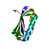
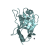
| Structure viewer | Molecule: MolmilJmol/JSmol |
|---|
- Downloads & links
-Download
| PDBx/mmCIF format | 4s1z.cif.gz | 105.8 KB | Display | PDBx/mmCIF format |
|---|---|---|---|---|
| PDB format | pdb4s1z.ent.gz | 79.5 KB | Display | PDB format |
| PDBx/mmJSON format | 4s1z.json.gz | Tree view | PDBx/mmJSON format | |
| Others |  Other downloads Other downloads |
-Validation report
| Summary document | 4s1z_validation.pdf.gz | 477 KB | Display | wwPDB validaton report |
|---|---|---|---|---|
| Full document | 4s1z_full_validation.pdf.gz | 478.1 KB | Display | |
| Data in XML | 4s1z_validation.xml.gz | 17.4 KB | Display | |
| Data in CIF | 4s1z_validation.cif.gz | 24.2 KB | Display | |
| Arichive directory | https://data.pdbj.org/pub/pdb/validation_reports/s1/4s1zftp://data.pdbj.org/pub/pdb/validation_reports/s1/4s1z | HTTPS FTP |
-Related structure data
| Related structure data |  4s22C  1ubqS  2wwzS C: citing same article ( S: Starting model for refinement |
|---|---|
| Similar structure data |


-Links
 PDBj
PDBj


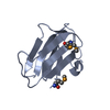
- Assembly
| Deposited unit | 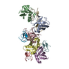
| ||||||||
|---|---|---|---|---|---|---|---|---|---|
| 1 | 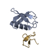
| ||||||||
| 2 | 
| ||||||||
| 3 | 
| ||||||||
| 4 | 
| ||||||||
| 5 | 
| ||||||||
| Unit cell |
|
-Components
| #1: Protein | Mass: 8576.831 Da / Num. of mol.: 5 / Fragment: residues 1-76 Source method: isolated from a genetically manipulated source Source: (gene. exp.) Homo sapiens (human) / Gene: UBA52, UBCEP2, ZRANB1 / Plasmid: pGEX6P1 / Production host:  #2: Protein/peptide | Mass: 4098.710 Da / Num. of mol.: 5 / Fragment: RanBP2-type 1 zinc finger domain residues 2-33 / Source method: isolated from a natural source / Source: (natural) #3: Chemical | ChemComp-ZN /   Mass: 65.409 Da / Num. of mol.: 5 / Source method: obtained synthetically / Formula: Zn Mass: 65.409 Da / Num. of mol.: 5 / Source method: obtained synthetically / Formula: Zn |
|---|
-Experimental details
-Experiment
| Experiment | Method: X-RAY DIFFRACTION / Number of used crystals: 1 |
|---|
- Sample preparation
| Crystal | Density Matthews: 3.69 Å3/Da / Density % sol: 66.68 % |
|---|---|
| Crystal grow | Temperature: 285 K / pH: 6.5 Details: 100mM MES, 200mM potassium iodide and 25% PEG4000, pH 6.5, VAPOR DIFFUSION, HANGING DROP, temperature 285K |
-Data collection
| Diffraction | Mean temperature: 285 K |
|---|---|
| Diffraction source | Source: SYNCHROTRON / Site: Diamond  / Beamline: I04 / Wavelength: 0.9795 / Beamline: I04 / Wavelength: 0.9795 |
| Detector | Type: DECTRIS PILATUS 6M-F / Detector: PIXEL / Date: Aug 10, 2014 / Details: COMPOUND REFRACTIVE LENSES |
| Radiation | Monochromator: DOUBLE CRYSTAL / Protocol: SINGLE WAVELENGTH / Monochromatic (M) / Laue (L): M / Scattering type: x-ray |
| Radiation wavelength | Wavelength: 0.9795 Å / Relative weight: 1 |
| Reflection | Resolution: 3→76.1 Å / Num. obs: 17755 / % possible obs: 99 % / Observed criterion σ(I): -3 / Redundancy: 3.5 % / Biso Wilson estimate: 85.71 Å2 / Rmerge(I) obs: 0.072 / Net I/σ(I): 15.64 |
| Reflection shell | Resolution: 3→3.1 Å / Redundancy: 3.4 % / Rmerge(I) obs: 0.4869 / Mean I/σ(I) obs: 2.05 / % possible all: 95.3 |
- Processing
| Software |
| ||||||||||||||||||||||||||||||||||||||||||||||||||||||||||||||||||||||||||||||||||||||||||||||||||||||||||||||||||||||||||||||||||||||||||||||||||||||||||||||||||||||||||||||||||||||
|---|---|---|---|---|---|---|---|---|---|---|---|---|---|---|---|---|---|---|---|---|---|---|---|---|---|---|---|---|---|---|---|---|---|---|---|---|---|---|---|---|---|---|---|---|---|---|---|---|---|---|---|---|---|---|---|---|---|---|---|---|---|---|---|---|---|---|---|---|---|---|---|---|---|---|---|---|---|---|---|---|---|---|---|---|---|---|---|---|---|---|---|---|---|---|---|---|---|---|---|---|---|---|---|---|---|---|---|---|---|---|---|---|---|---|---|---|---|---|---|---|---|---|---|---|---|---|---|---|---|---|---|---|---|---|---|---|---|---|---|---|---|---|---|---|---|---|---|---|---|---|---|---|---|---|---|---|---|---|---|---|---|---|---|---|---|---|---|---|---|---|---|---|---|---|---|---|---|---|---|---|---|---|---|
| Refinement | Method to determine structure: MOLECULAR REPLACEMENT Starting model: 2WWZ, 1UBQ Resolution: 3.03→76.1 Å / Cor.coef. Fo:Fc: 0.939 / Cor.coef. Fo:Fc free: 0.909 / SU B: 22.303 / SU ML: 0.382 / Cross valid method: THROUGHOUT / σ(F): 0 / ESU R: 0.866 / ESU R Free: 0.389 / Stereochemistry target values: MAXIMUM LIKELIHOOD / Details: HYDROGENS HAVE BEEN ADDED IN THE RIDING POSITIONS
| ||||||||||||||||||||||||||||||||||||||||||||||||||||||||||||||||||||||||||||||||||||||||||||||||||||||||||||||||||||||||||||||||||||||||||||||||||||||||||||||||||||||||||||||||||||||
| Solvent computation | Ion probe radii: 0.8 Å / Shrinkage radii: 0.8 Å / VDW probe radii: 1.2 Å / Solvent model: MASK | ||||||||||||||||||||||||||||||||||||||||||||||||||||||||||||||||||||||||||||||||||||||||||||||||||||||||||||||||||||||||||||||||||||||||||||||||||||||||||||||||||||||||||||||||||||||
| Displacement parameters | Biso mean: 81.9 Å2
| ||||||||||||||||||||||||||||||||||||||||||||||||||||||||||||||||||||||||||||||||||||||||||||||||||||||||||||||||||||||||||||||||||||||||||||||||||||||||||||||||||||||||||||||||||||||
| Refinement step | Cycle: LAST / Resolution: 3.03→76.1 Å
| ||||||||||||||||||||||||||||||||||||||||||||||||||||||||||||||||||||||||||||||||||||||||||||||||||||||||||||||||||||||||||||||||||||||||||||||||||||||||||||||||||||||||||||||||||||||
| Refine LS restraints |
|