 Movie
Movie Controller

[English] 日本語
 Yorodumi
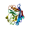
- PDB-4s1e: Crystal structure of cyclophilin mutant L120A from Leishmania don... -
+ Open data

- Basic information
| Entry | Database: PDB / ID: 4s1e | ||||||
|---|---|---|---|---|---|---|---|
| Title | Crystal structure of cyclophilin mutant L120A from Leishmania donovani at 2.22 angstrom. | ||||||
 Components Components | Peptidyl-prolyl cis-trans isomerase | ||||||
 Keywords Keywords | ISOMERASE / Cytosol | ||||||
| Function / homology |  Function and homology information Function and homology informationcyclosporin A binding / peptidylprolyl isomerase / peptidyl-prolyl cis-trans isomerase activity / protein folding / cytoplasm Similarity search - Function | ||||||
| Biological species |  Leishmania donovani (eukaryote) Leishmania donovani (eukaryote) | ||||||
| Method |  X-RAY DIFFRACTION / MOLECULAR REPLACEMENT / Resolution: 2.22 Å X-RAY DIFFRACTION / MOLECULAR REPLACEMENT / Resolution: 2.22 Å | ||||||
 Authors Authors | Roy, S. / Datta, A.K. / Banerjee, R. | ||||||
 Citation Citation | Journal: To be Published Title: Characterization and prediction of thermal stability of cyclophilin mutants from L.donovani Authors: Roy, S. / Datta, A.K. / Banerjee, R. | ||||||
| History |
|
- Structure visualization
| Structure viewer | Molecule: MolmilJmol/JSmol |
|---|
- Downloads & links
-Download
| PDBx/mmCIF format | 4s1e.cif.gz | 77.1 KB | Display | PDBx/mmCIF format |
|---|---|---|---|---|
| PDB format | pdb4s1e.ent.gz | 57.4 KB | Display | PDB format |
| PDBx/mmJSON format | 4s1e.json.gz | Tree view | PDBx/mmJSON format | |
| Others |  Other downloads Other downloads |
-Validation report
| Arichive directory | https://data.pdbj.org/pub/pdb/validation_reports/s1/4s1eftp://data.pdbj.org/pub/pdb/validation_reports/s1/4s1e | HTTPS FTP |
|---|
-Related structure data
| Related structure data |  4s1jC  2haqS S: Starting model for refinement C: citing same article ( |
|---|---|
| Similar structure data |


-Links
 PDBj
PDBj

- Assembly
| Deposited unit | 
| ||||||||
|---|---|---|---|---|---|---|---|---|---|
| 1 | 
| ||||||||
| 2 | 
| ||||||||
| Unit cell |
|
-Components
| #1: Protein | Mass: 19032.525 Da / Num. of mol.: 2 / Fragment: residues 22-187 / Mutation: L120A Source method: isolated from a genetically manipulated source Source: (gene. exp.) Leishmania donovani (eukaryote) / Gene: CYP / Plasmid: pQE32 / Production host:  #2: Water | ChemComp-HOH / |  Mass: 18.015 Da / Num. of mol.: 119 / Source method: isolated from a natural source / Formula: H2O Mass: 18.015 Da / Num. of mol.: 119 / Source method: isolated from a natural source / Formula: H2OSequence details | THE DEPOSITOR STATES THAT THE PRIMARY SEQUENCE WAS RE-SEQUENCED AND ERRORS WERE DETECTED AT ...THE DEPOSITOR STATES THAT THE PRIMARY SEQUENCE WAS RE-SEQUENCED AND ERRORS WERE DETECTED AT POSITIONS 81 AND 112. THE ACTUAL SEQUENCE WAS FOUND TO BE PRO 81 AND LYS 112, WHICH WAS ALSO CONFIRMED IN THE ELECTRON DENSITY MAP OF THE STRUCTURE. | |
|---|
-Experimental details
-Experiment
| Experiment | Method: X-RAY DIFFRACTION / Number of used crystals: 1 |
|---|
- Sample preparation
| Crystal | Density Matthews: 2.19 Å3/Da / Density % sol: 43.83 % |
|---|---|
| Crystal grow | Temperature: 298 K / Method: vapor diffusion, hanging drop / pH: 8 Details: 10% PEG 3350,0.02 M Tris-HCl,0.02% Azide, pH 8, VAPOR DIFFUSION, HANGING DROP, temperature 298.0K |
-Data collection
| Diffraction | Mean temperature: 298 K | |||||||||||||||||||||
|---|---|---|---|---|---|---|---|---|---|---|---|---|---|---|---|---|---|---|---|---|---|---|
| Diffraction source | Source: ROTATING ANODE / Type: RIGAKU RU200 / Wavelength: 1.5418 Å | |||||||||||||||||||||
| Detector | Type: MAR scanner 300 mm plate / Detector: IMAGE PLATE / Date: Sep 12, 2011 | |||||||||||||||||||||
| Radiation | Monochromator: Mirrors / Protocol: SINGLE WAVELENGTH / Monochromatic (M) / Laue (L): M / Scattering type: x-ray | |||||||||||||||||||||
| Radiation wavelength | Wavelength: 1.5418 Å / Relative weight: 1 | |||||||||||||||||||||
| Reflection | Resolution: 2.22→30 Å / Num. all: 16104 / Num. obs: 16104 / % possible obs: 99.2 % / Observed criterion σ(F): 0 / Observed criterion σ(I): 0 / Redundancy: 5.91 % / Biso Wilson estimate: 32.5 Å2 / Rmerge(I) obs: 0.056 / Net I/σ(I): 23.8 | |||||||||||||||||||||
| Reflection shell |
|
- Processing
| Software |
| ||||||||||||||||||||
|---|---|---|---|---|---|---|---|---|---|---|---|---|---|---|---|---|---|---|---|---|---|
| Refinement | Method to determine structure: MOLECULAR REPLACEMENT Starting model: 2HAQ Resolution: 2.22→30 Å / Isotropic thermal model: Isotropic / Cross valid method: THROUGHOUT / σ(F): 1 / Stereochemistry target values: Engh & Huber
| ||||||||||||||||||||
| Displacement parameters | Biso mean: 30.85 Å2
| ||||||||||||||||||||
| Refinement step | Cycle: LAST / Resolution: 2.22→30 Å
| ||||||||||||||||||||
| Refine LS restraints |
|
