 Movie
Movie Controller


+ Open data

- Basic information
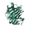
| Entry | Database: PDB / ID: 2haq | ||||||
|---|---|---|---|---|---|---|---|
| Title | Crystal Structure of Cyclophilin A from Leishmania Donovani | ||||||
 Components Components | Cyclophilin | ||||||
 Keywords Keywords | ISOMERASE / CYCLOPHILIN / ROTAMASE / PROLINE / CIS-TRANS / PROTOZOA / LEISHMANIA / DONOVANI / KALA-AZAR. | ||||||
| Function / homology |  Function and homology information Function and homology informationcyclosporin A binding / peptidylprolyl isomerase / peptidyl-prolyl cis-trans isomerase activity / protein folding / cytoplasm Similarity search - Function | ||||||
| Biological species |  Leishmania donovani (eukaryote) Leishmania donovani (eukaryote) | ||||||
| Method |  X-RAY DIFFRACTION / MOLECULAR REPLACEMENT / Resolution: 1.97 Å X-RAY DIFFRACTION / MOLECULAR REPLACEMENT / Resolution: 1.97 Å | ||||||
 Authors Authors | Venugopal, V. / Sen, B. / Datta, A.K. / Banerjee, R. | ||||||
 Citation Citation | Journal: Acta Crystallogr.,Sect.F / Year: 2007 Title: Structure of cyclophilin from Leishmania donovani at 1.97 A resolution. Authors: Venugopal, V. / Sen, B. / Datta, A.K. / Banerjee, R. #1: Journal: J.Biol.Chem. / Year: 2001Title: Lack of Abundance of Cytoplasmic Cyclosporin A Bind Protein Renders Free Living Leishmania Donovani Resistant to Cyclosporin A Authors: Dutta, M. / Delhi, P. / Sinha, K.M. / Banerjee, R. / Datta, A.K. #2: Journal: Acta Crystallogr.,Sect.D / Year: 2002Title: Crystallisation and Preliminary X-Ray Analysis of Cyclophilin from Leishmania Donovani Authors: Banerjee, R. / Dutta, M. / Sen, M. / Datta, A.K. #3: Journal: Curr.Sci. / Year: 2004Title: Crystal Structure of Cyclophilin from Leishmania Donovani at 3.5 A Resolution Authors: Banerjee, R. / Dutta, M. / Sen, M. / Datta, A.K. | ||||||
| History |
|
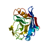
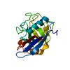
- Structure visualization
| Structure viewer | Molecule: MolmilJmol/JSmol |
|---|
- Downloads & links
-Download
| PDBx/mmCIF format | 2haq.cif.gz | 47.6 KB | Display | PDBx/mmCIF format |
|---|---|---|---|---|
| PDB format | pdb2haq.ent.gz | 32.7 KB | Display | PDB format |
| PDBx/mmJSON format | 2haq.json.gz | Tree view | PDBx/mmJSON format | |
| Others |  Other downloads Other downloads |
-Validation report
| Arichive directory | https://data.pdbj.org/pub/pdb/validation_reports/ha/2haqftp://data.pdbj.org/pub/pdb/validation_reports/ha/2haq | HTTPS FTP |
|---|
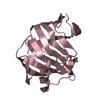
-Related structure data
| Related structure data |  1xo7S S: Starting model for refinement |
|---|---|
| Similar structure data |


-Links
 PDBj
PDBj

- Assembly
| Deposited unit | 
| ||||||||
|---|---|---|---|---|---|---|---|---|---|
| 1 |
| ||||||||
| Unit cell |
| ||||||||
| Details | The protein is a monomer |
-Components
| #1: Protein | Mass: 19090.541 Da / Num. of mol.: 1 Source method: isolated from a genetically manipulated source Source: (gene. exp.) Leishmania donovani (eukaryote) / Gene: CYP / Plasmid: pQE32 / Production host:  |
|---|---|
| #2: Water | ChemComp-HOH /  Mass: 18.015 Da / Num. of mol.: 102 / Source method: isolated from a natural source / Formula: H2O Mass: 18.015 Da / Num. of mol.: 102 / Source method: isolated from a natural source / Formula: H2O |
-Experimental details
-Experiment
| Experiment | Method: X-RAY DIFFRACTION / Number of used crystals: 1 |
|---|
- Sample preparation
| Crystal | Density Matthews: 2.3 Å3/Da / Density % sol: 45.6 % |
|---|---|
| Crystal grow | Temperature: 293 K / Method: vapor diffusion, hanging drop / pH: 8.5 Details: 0.01M TRIS, 7.5% PEG3350, 0.005M IMID 0.02% AZIDE, PH 8.5, VAPOR DIFFUSION, HANGING DROP, TEMPERATURE RESERVOIR CONTAINING 40% PEG3350 IN THE SAME BUFFER. PROTEIN CONCENTRATION: 10 MG/ML, temperature 293K |
-Data collection
| Diffraction | Mean temperature: 120 K |
|---|---|
| Diffraction source | Source: ROTATING ANODE / Type: RIGAKU RU200 / Wavelength: 1.5418 Å |
| Detector | Type: MAR scanner 300 mm plate / Detector: IMAGE PLATE / Date: Jun 8, 2004 / Details: OSMIC MAXFLUX CONFOCAL MULTILAY |
| Radiation | Monochromator: Mirrors / Protocol: SINGLE WAVELENGTH / Monochromatic (M) / Laue (L): M / Scattering type: x-ray |
| Radiation wavelength | Wavelength: 1.5418 Å / Relative weight: 1 |
| Reflection | Resolution: 1.97→14.93 Å / Num. all: 12644 / Num. obs: 12516 / % possible obs: 99 % / Observed criterion σ(F): 1 / Observed criterion σ(I): 1 / Redundancy: 8.1 % / Biso Wilson estimate: 24.4 Å2 / Rmerge(I) obs: 0.068 / Net I/σ(I): 30.1 |
| Reflection shell | Resolution: 1.97→2.02 Å / Redundancy: 5.2 % / Rmerge(I) obs: 0.293 / Mean I/σ(I) obs: 4.5 / % possible all: 86.6 |
- Processing
| Software |
| |||||||||||||||||||||||||
|---|---|---|---|---|---|---|---|---|---|---|---|---|---|---|---|---|---|---|---|---|---|---|---|---|---|---|
| Refinement | Method to determine structure: MOLECULAR REPLACEMENT Starting model: 1XO7.PDB Resolution: 1.97→14.93 Å / Rfactor Rfree error: 0.007 / Isotropic thermal model: RESTRAINED / Cross valid method: THROUGHOUT / σ(F): 2 / Stereochemistry target values: Engh & Huber
| |||||||||||||||||||||||||
| Displacement parameters | Biso mean: 28.5 Å2
| |||||||||||||||||||||||||
| Refine analyze |
| |||||||||||||||||||||||||
| Refinement step | Cycle: LAST / Resolution: 1.97→14.93 Å
| |||||||||||||||||||||||||
| Refine LS restraints |
| |||||||||||||||||||||||||
| LS refinement shell | Resolution: 1.97→2.09 Å / Rfactor Rfree error: 0.022 / Total num. of bins used: 6
| |||||||||||||||||||||||||
| Xplor file |
|
