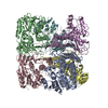
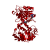
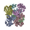
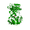
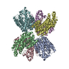
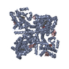
[English] 日本語
Yorodumi
- PDB-4s14: Crystal structure of the orphan nuclear receptor RORgamma ligand-... -

+
Open data


ID or keywords:

Loading...

-
Basic information

Entry
Database: PDB / ID: 4s14
TitleCrystal structure of the orphan nuclear receptor RORgamma ligand-binding domain in complex with 4alpha-caboxyl, 4beta-methyl-zymosterol (4ACD8)
Components
  • Nuclear receptor ROR-gamma
  • Nuclear receptor-interacting protein 1
KeywordsTRANSCRIPTION / transcription factor
Function / homology
Function and homology information


ovarian follicle rupture / NR1H2 & NR1H3 regulate gene expression linked to gluconeogenesis / T-helper 17 cell differentiation / ligand-activated transcription factor activity / cellular response to sterol / regulation of steroid metabolic process / NR1H2 & NR1H3 regulate gene expression linked to lipogenesis / lipid storage / nuclear glucocorticoid receptor binding / Peyer's patch development ...ovarian follicle rupture / NR1H2 & NR1H3 regulate gene expression linked to gluconeogenesis / T-helper 17 cell differentiation / ligand-activated transcription factor activity / cellular response to sterol / regulation of steroid metabolic process / NR1H2 & NR1H3 regulate gene expression linked to lipogenesis / lipid storage / nuclear glucocorticoid receptor binding / Peyer's patch development / positive regulation of circadian rhythm / oxysterol binding / T-helper cell differentiation / RUNX3 Regulates Immune Response and Cell Migration / negative regulation of thymocyte apoptotic process / regulation of fat cell differentiation / histone deacetylase complex / regulation of glucose metabolic process / lymph node development / adipose tissue development / nuclear retinoid X receptor binding / xenobiotic metabolic process / SUMOylation of transcription cofactors / cellular response to estradiol stimulus / nuclear estrogen receptor binding / nuclear receptor binding / circadian regulation of gene expression / Heme signaling / fibrillar center / DNA-binding transcription repressor activity, RNA polymerase II-specific / histone deacetylase binding / Nuclear Receptor transcription pathway / circadian rhythm / transcription corepressor activity / nuclear receptor activity / sequence-specific double-stranded DNA binding / Circadian Clock / Interleukin-4 and Interleukin-13 signaling / Estrogen-dependent gene expression / transcription coactivator activity / nuclear body / DNA-binding transcription factor activity, RNA polymerase II-specific / nuclear speck / RNA polymerase II cis-regulatory region sequence-specific DNA binding / DNA-binding transcription factor activity / signaling receptor binding / chromatin / nucleolus / regulation of transcription by RNA polymerase II / positive regulation of DNA-templated transcription / negative regulation of transcription by RNA polymerase II / positive regulation of transcription by RNA polymerase II / zinc ion binding / nucleoplasm / nucleus / cytosol
Similarity search - Function
Nuclear receptor-interacting protein 1 / Nuclear receptor-interacting protein 1, repression domain 1 / Nuclear receptor-interacting protein 1, repression domain 2 / Nuclear receptor-interacting protein 1, repression domain 3 / Nuclear receptor-interacting protein 1, repression domain 4 / Nuclear receptor-interacting protein 1 repression 1 / Nuclear receptor-interacting protein 1 repression 2 / Nuclear receptor-interacting protein 1 repression 3 / Nuclear receptor-interacting protein 1 repression 4 / Nuclear receptor ROR ...Nuclear receptor-interacting protein 1 / Nuclear receptor-interacting protein 1, repression domain 1 / Nuclear receptor-interacting protein 1, repression domain 2 / Nuclear receptor-interacting protein 1, repression domain 3 / Nuclear receptor-interacting protein 1, repression domain 4 / Nuclear receptor-interacting protein 1 repression 1 / Nuclear receptor-interacting protein 1 repression 2 / Nuclear receptor-interacting protein 1 repression 3 / Nuclear receptor-interacting protein 1 repression 4 / Nuclear receptor ROR / Retinoid-related orphan receptors, DNA-binding domain / Retinoid X Receptor / Retinoid X Receptor / Nuclear hormone receptor / Nuclear hormones receptors DNA-binding region signature. / Zinc finger, nuclear hormone receptor-type / Zinc finger, C4 type (two domains) / Nuclear hormone receptors DNA-binding domain profile. / c4 zinc finger in nuclear hormone receptors / Nuclear hormone receptor, ligand-binding domain / Nuclear hormone receptor-like domain superfamily / Ligand-binding domain of nuclear hormone receptor / Nuclear receptor (NR) ligand-binding (LBD) domain profile. / Ligand binding domain of hormone receptors / Zinc finger, NHR/GATA-type / Orthogonal Bundle / Mainly Alpha
Similarity search - Domain/homology
Chem-4D8 / Nuclear receptor-interacting protein 1 / Nuclear receptor ROR-gamma
Similarity search - Component
Biological speciesHomo sapiens (human)
MethodX-RAY DIFFRACTION / SYNCHROTRON / MOLECULAR REPLACEMENT / Resolution: 3.542 Å
AuthorsHuang, P. / Santori, F.R. / Littman, D.R. / Rastinejad, F.
CitationJournal: Cell Metab / Year: 2015
Title: Identification of Natural ROR gamma Ligands that Regulate the Development of Lymphoid Cells.
Authors: Santori, F.R. / Huang, P. / van de Pavert, S.A. / Douglass, E.F. / Leaver, D.J. / Haubrich, B.A. / Keber, R. / Lorbek, G. / Konijn, T. / Rosales, B.N. / Rozman, D. / Horvat, S. / Rahier, A. ...Authors: Santori, F.R. / Huang, P. / van de Pavert, S.A. / Douglass, E.F. / Leaver, D.J. / Haubrich, B.A. / Keber, R. / Lorbek, G. / Konijn, T. / Rosales, B.N. / Rozman, D. / Horvat, S. / Rahier, A. / Mebius, R.E. / Rastinejad, F. / Nes, W.D. / Littman, D.R.
History
DepositionJan 7, 2015Deposition site: RCSB / Processing site: RCSB
Revision 1.0Feb 11, 2015Provider: repository / Type: Initial release
Revision 1.1Feb 25, 2015Group: Database references

-
Structure visualization

Structure viewerMolecule:
MolmilJmol/JSmol

Downloads & links

-
Assembly

Deposited unit
A: Nuclear receptor ROR-gamma
C: Nuclear receptor-interacting protein 1
hetero molecules


Theoretical massNumber of molelcules
Total (without water)31,7373
Polymers31,2942
Non-polymers4431
Water00
1
A: Nuclear receptor ROR-gamma
C: Nuclear receptor-interacting protein 1
hetero molecules
x 6


Theoretical massNumber of molelcules
Total (without water)190,42118
Polymers187,76512
Non-polymers2,6566
Water0
TypeNameSymmetry operationNumber
identity operation1_555x,y,z1
crystal symmetry operation2_545-y,x-y-1,z1
crystal symmetry operation3_655-x+y+1,-x,z1
crystal symmetry operation10_554-y,-x,-z-1/21
crystal symmetry operation11_654-x+y+1,y,-z-1/21
crystal symmetry operation12_544x,x-y-1,-z-1/21
Buried area14880 Å2
ΔGint-81 kcal/mol
Surface area65420 Å2
MethodPISA
2


  • Idetical with deposited unit
  • defined by author&software
TypeNameSymmetry operationNumber
identity operation1_555x,y,z1
Buried area910 Å2
ΔGint-8 kcal/mol
Surface area12470 Å2
MethodPISA
Unit cell
Length a, b, c (Å)158.179, 158.179, 129.801
Angle α, β, γ (deg.)90.00, 90.00, 120.00
Int Tables number182
Space group name H-MP6322

-
Components

#1: Protein Nuclear receptor ROR-gamma / Nuclear receptor RZR-gamma / Nuclear receptor subfamily 1 group F member 3 / RAR-related orphan ...Nuclear receptor RZR-gamma / Nuclear receptor subfamily 1 group F member 3 / RAR-related orphan receptor C / Retinoid-related orphan receptor-gamma


Mass: 29913.531 Da / Num. of mol.: 1 / Fragment: ligand-binding domain
Source method: isolated from a genetically manipulated source
Source: (gene. exp.) Homo sapiens (human) / Gene: NR1F3, RORC, RORG, RZRG / Plasmid: pET46 / Production host: Escherichia coli (E. coli) / Strain (production host): BL21-CodonPlus(DE3)-RIL / References: UniProt: P51449
#2: Protein/peptide Nuclear receptor-interacting protein 1 / Nuclear factor RIP140 / Receptor-interacting protein 140


Mass: 1380.632 Da / Num. of mol.: 1 / Fragment: LxxLL binding motif / Source method: obtained synthetically / Details: This sequence occurs naturally in humans. / Source: (synth.) Homo sapiens (human) / References: UniProt: P48552
#3: Chemical ChemComp-4D8 / (3beta,4alpha,5beta,14beta)-3-hydroxy-4-methylcholesta-8,24-diene-4-carboxylic acid / 4alpha-carboxy-4beta-methyl-zymosterol


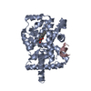
Mass: 442.674 Da / Num. of mol.: 1 / Source method: obtained synthetically / Formula: C29H46O3

-
Experimental details

-
Experiment

ExperimentMethod: X-RAY DIFFRACTION / Number of used crystals: 1

-
Sample preparation

Crystal growTemperature: 282 K / Method: vapor diffusion, sitting drop / pH: 8
Details: 0.3M Potassium sodium tartrate, pH 8, VAPOR DIFFUSION, SITTING DROP, temperature 282K

-
Data collection

DiffractionMean temperature: 100 K
Diffraction sourceSource: SYNCHROTRON / Site: APS / Beamline: 19-ID / Wavelength: 0.9793 Å
DetectorType: ADSC QUANTUM 315r / Detector: CCD / Date: Jul 1, 2012
RadiationMonochromator: Rosenbaum-Rock high-resolution double-crystal Si(111)
Protocol: SINGLE WAVELENGTH / Monochromatic (M) / Laue (L): M / Scattering type: x-ray
Radiation wavelengthWavelength: 0.9793 Å / Relative weight: 1
ReflectionResolution: 3.54→50 Å / Num. all: 12173 / Num. obs: 12173 / % possible obs: 100 % / Observed criterion σ(F): 0 / Observed criterion σ(I): 0 / Redundancy: 11.7 % / Net I/σ(I): 16

-
Processing

Software
NameVersionClassification
HKL-3000data collection
PHASERphasing
PHENIX(phenix.refine: 1.8.2_1309)refinement
HKL-3000data reduction
HKL-3000data scaling
RefinementMethod to determine structure: MOLECULAR REPLACEMENT / Resolution: 3.542→47.111 Å / SU ML: 0.32 / σ(F): 1.35 / Phase error: 19.57 / Stereochemistry target values: ML
RfactorNum. reflection% reflectionSelection details
Rfree0.2193 567 4.71 %RANDOM
Rwork0.1867 ---
all0.1882 12156 --
obs0.1882 12041 99.05 %-
Solvent computationShrinkage radii: 0.9 Å / VDW probe radii: 1.11 Å / Solvent model: FLAT BULK SOLVENT MODEL
Refinement stepCycle: LAST / Resolution: 3.542→47.111 Å
ProteinNucleic acidLigandSolventTotal
Num. atoms2075 0 32 0 2107
Refine LS restraints
Refine-IDTypeDev idealNumber
X-RAY DIFFRACTIONf_bond_d0.0032156
X-RAY DIFFRACTIONf_angle_d0.7552912
X-RAY DIFFRACTIONf_dihedral_angle_d15.244817
X-RAY DIFFRACTIONf_chiral_restr0.048324
X-RAY DIFFRACTIONf_plane_restr0.002366
LS refinement shell
Resolution (Å)Rfactor RfreeNum. reflection RfreeRfactor RworkNum. reflection RworkRefine-ID% reflection obs (%)
3.5418-3.89810.22841410.20562765X-RAY DIFFRACTION98
3.8981-4.46180.23091460.17382816X-RAY DIFFRACTION99
4.4618-5.61990.18571520.17072851X-RAY DIFFRACTION99
5.6199-47.11470.23641280.1983042X-RAY DIFFRACTION99
Refinement TLS params.

Method: refined / Refine-ID: X-RAY DIFFRACTION

IDL112)L122)L132)L222)L232)L332)S11 (Å °)S12 (Å °)S13 (Å °)S21 (Å °)S22 (Å °)S23 (Å °)S31 (Å °)S32 (Å °)S33 (Å °)T112)T122)T132)T222)T232)T332)Origin x (Å)Origin y (Å)Origin z (Å)
13.21591.124-2.22821.7597-0.97442.52580.0961-0.11220.10560.30150.08550.43810.1623-0.28-0.12530.4956-0.06670.14070.47690.13530.506951.1448-45.2917-14.6793
22.32561.60161.24612.5738-0.66763.82990.0829-0.33140.20940.1524-0.2190.0306-0.22380.0637-0.41340.7155-0.14010.31130.98-0.08280.534353.8567-35.41632.4844
Refinement TLS group
IDRefine-IDRefine TLS-IDSelection details
1X-RAY DIFFRACTION1chain A
2X-RAY DIFFRACTION2chain C

+
About Yorodumi

-
News

-
Feb 9, 2022. New format data for meta-information of EMDB entries

New format data for meta-information of EMDB entries

  • Version 3 of the EMDB header file is now the official format.
  • The previous official version 1.9 will be removed from the archive.

Related info.:EMDB header

External links:wwPDB to switch to version 3 of the EMDB data model

-
Aug 12, 2020. Covid-19 info

Covid-19 info

URL: https://pdbjlvh1.pdbj.org/emnavi/covid19.php

New page: Covid-19 featured information page in EM Navigator.

Related info.:Covid-19 info / Mar 5, 2020. Novel coronavirus structure data

+
Mar 5, 2020. Novel coronavirus structure data

Novel coronavirus structure data

Related info.:Yorodumi Speices / Aug 12, 2020. Covid-19 info

External links:COVID-19 featured content - PDBj / Molecule of the Month (242):Coronavirus Proteases

+
Jan 31, 2019. EMDB accession codes are about to change! (news from PDBe EMDB page)

EMDB accession codes are about to change! (news from PDBe EMDB page)

  • The allocation of 4 digits for EMDB accession codes will soon come to an end. Whilst these codes will remain in use, new EMDB accession codes will include an additional digit and will expand incrementally as the available range of codes is exhausted. The current 4-digit format prefixed with “EMD-” (i.e. EMD-XXXX) will advance to a 5-digit format (i.e. EMD-XXXXX), and so on. It is currently estimated that the 4-digit codes will be depleted around Spring 2019, at which point the 5-digit format will come into force.
  • The EM Navigator/Yorodumi systems omit the EMD- prefix.

Related info.:Q: What is EMD? / ID/Accession-code notation in Yorodumi/EM Navigator

External links:EMDB Accession Codes are Changing Soon! / Contact to PDBj

+
Jul 12, 2017. Major update of PDB

Major update of PDB

  • wwPDB released updated PDB data conforming to the new PDBx/mmCIF dictionary.
  • This is a major update changing the version number from 4 to 5, and with Remediation, in which all the entries are updated.
  • In this update, many items about electron microscopy experimental information are reorganized (e.g. em_software).
  • Now, EM Navigator and Yorodumi are based on the updated data.

External links:wwPDB Remediation / Enriched Model Files Conforming to OneDep Data Standards Now Available in the PDB FTP Archive

-
Yorodumi

Thousand views of thousand structures

  • Yorodumi is a browser for structure data from EMDB, PDB, SASBDB, etc.
  • This page is also the successor to EM Navigator detail page, and also detail information page/front-end page for Omokage search.
  • The word "yorodu" (or yorozu) is an old Japanese word meaning "ten thousand". "mi" (miru) is to see.

Related info.:EMDB / PDB / SASBDB / Comparison of 3 databanks / Yorodumi Search / Aug 31, 2016. New EM Navigator & Yorodumi / Yorodumi Papers / Jmol/JSmol / Function and homology information / Changes in new EM Navigator and Yorodumi

Read more